


Happy Holidays! All of this week’s posts will be themed around the winter holiday season. Today’s path rounds are on 𝐫𝐞𝐝 𝐧𝐨𝐬𝐞!
𝐖𝐡𝐚𝐭 𝐢𝐬 𝐢𝐭?
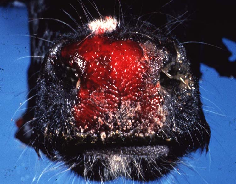
𝐑𝐞𝐝 𝐧𝐨𝐬𝐞 is more scientifically known as 𝐢𝐧𝐟𝐞𝐜𝐭𝐢𝐨𝐮𝐬 𝐛𝐨𝐯𝐢𝐧𝐞 𝐫𝐡𝐢𝐧𝐨𝐭𝐫𝐚𝐜𝐡𝐞𝐢𝐭𝐢𝐬, and is a viral infection affecting the upper respiratory tract. As the name might suggest, affected cattle often have a 𝐡𝐲𝐩𝐞𝐫𝐞𝐦𝐢𝐜 (red) nose!
𝐖𝐡𝐨 𝐠𝐞𝐭𝐬 𝐢𝐭?
This disease is typically found in feedlot cattle.
𝐖𝐡𝐚𝐭 𝐜𝐚𝐮𝐬𝐞𝐬 𝐢𝐭?
Red nose is caused by 𝐛𝐨𝐯𝐢𝐧𝐞 𝐡𝐞𝐫𝐩𝐞𝐬𝐯𝐢𝐫𝐮𝐬-𝟏. Animals will inhale the virus through aerosols or direct contact with nasal secretions from infected cattle. From there, the virus covers the respiratory mucosa, and enters the 𝐭𝐫𝐢𝐠𝐞𝐦𝐢𝐧𝐚𝐥 𝐧𝐞𝐫𝐯𝐞, where it makes itself a little home. The virus can become 𝐥𝐚𝐭𝐞𝐧𝐭 (non-infectious but still present) here, and can cause repeated infections when the animal is stressed. This process is similar to cold sores in humans, which are also caused by a herpesvirus!
𝐖𝐡𝐲 𝐢𝐬 𝐭𝐡𝐢𝐬 𝐚 𝐩𝐫𝐨𝐛𝐥𝐞𝐦?
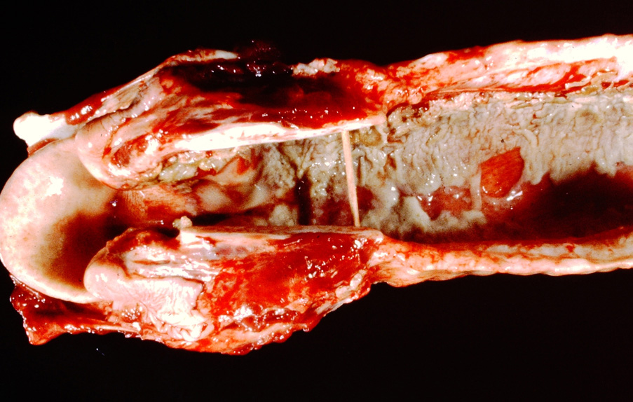
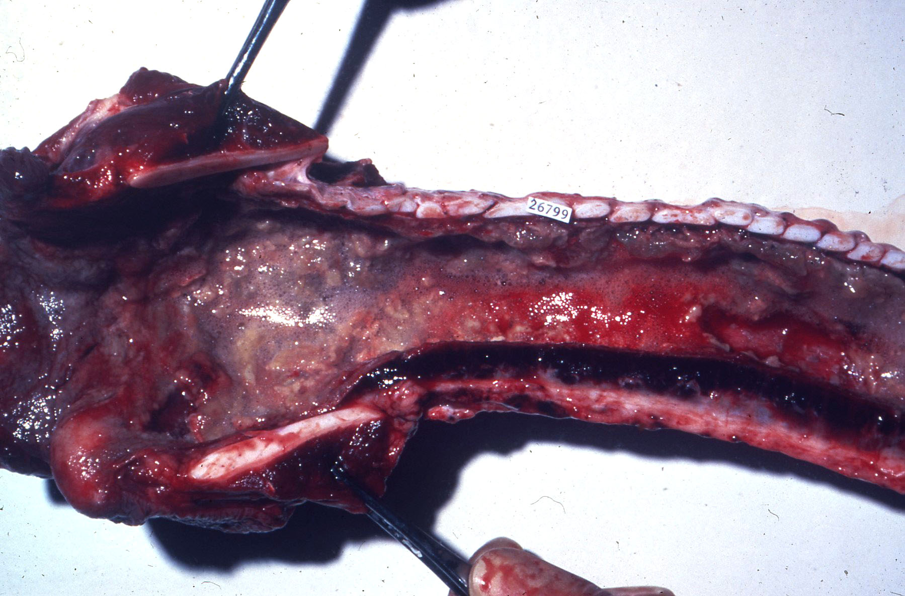
In its disease-causing form, the virus destroys the 𝐞𝐩𝐢𝐭𝐡𝐞𝐥𝐢𝐚𝐥 𝐜𝐞𝐥𝐥𝐬 (surface cells) of the nasal passages and airways. This causes 𝐞𝐫𝐨𝐬𝐢𝐨𝐧𝐬 to form, where the mucosal surface is completely lost. Because these tissues often have a lot of vasculature, the vessels under the erosions will secrete 𝐟𝐢𝐛𝐫𝐢𝐧, a major clotting protein, in order to try and fill in the defect. This fibrin ultimately combines with the dead epithelial cells producing a 𝐟𝐢𝐛𝐫𝐢𝐧𝐨𝐧𝐞𝐜𝐫𝐨𝐭𝐢𝐜 𝐦𝐞𝐦𝐛𝐫𝐚𝐧𝐞, which is characteristic of the disease.
In some cases, affected cattle can also develop a pneumonia, because the epithelial cells of the trachea are compromised. These epithelial cells have long 𝐜𝐢𝐥𝐢𝐚 (finger-like projections) that help push bacteria, foreign materials, and mucus back up the throat to be coughed out. Without these cilia, anything that ends up in the trachea falls right into the lungs, and can cause pneumonia.
𝐇𝐨𝐰 𝐢𝐬 𝐢𝐭 𝐝𝐢𝐚𝐠𝐧𝐨𝐬𝐞𝐝?
The veterinarian might suspect the disease based on a necropsy, or clinical signs of affected animals. From there, viral isolation can be conducted to confirm the presence of the virus.
𝐇𝐨𝐰 𝐢𝐬 𝐢𝐭 𝐭𝐫𝐞𝐚𝐭𝐞𝐝?
Because these cattle are predisposed to pneumonia, they often get a dose of antibiotics to either prevent a pneumonia, or treat an existing one. Eventually, the animal will likely recover with the virus going back to its latent state.
𝐇𝐨𝐰 𝐢𝐬 𝐢𝐭 𝐩𝐫𝐞𝐯𝐞𝐧𝐭𝐞𝐝?
Thankfully, this virus can be easily prevented using vaccines! Other recommendations include low-stress handling, avoiding mixing groups of cattle from different locations, and ensuring the animals have access to feed and water to lower stress.
𝐏𝐡𝐨𝐭𝐨𝐬
1) A cow with a red nose from IBR!
2-4) Examples of the fibrinonecrotic membrane in the trachea from IBR.
5) More fibrinonecrotic membranes, this time in the nasal passages!
𝐒𝐨𝐮𝐫𝐜𝐞𝐬
Maxie, G. Jubb, Kennedy and Palmer’s Pathology of Domestic Animals, Volume 2. Sixth Edition.
Photos 1-5 © Noah’s Arkive contributors Jakowski, Doster, King, Williams licensed under CC BY-SA 4.0.


