Today’s path rounds are on 𝐯𝐞𝐧𝐭𝐫𝐢𝐜𝐮𝐥𝐚𝐫 𝐬𝐞𝐩𝐭𝐚𝐥 𝐝𝐞𝐟𝐞𝐜𝐭𝐬! This topic inspired by the second year systemic pathology labs I helped out in this week ![]()
𝐖𝐡𝐚𝐭 𝐢𝐬 𝐢𝐭?
𝐕𝐞𝐧𝐭𝐫𝐢𝐜𝐮𝐥𝐚𝐫 𝐬𝐞𝐩𝐭𝐚𝐥 𝐝𝐞𝐟𝐞𝐜𝐭𝐬 are probably the most common developmental abnormality of the heart! In this disease, there is incomplete formation of the 𝐢𝐧𝐭𝐞𝐫𝐯𝐞𝐧𝐭𝐫𝐢𝐜𝐮𝐥𝐚𝐫 𝐬𝐞𝐩𝐭𝐮𝐦, which divides the two sides of the heart. 𝐖𝐡𝐨 𝐠𝐞𝐭𝐬 𝐢𝐭?Any species can get this condition!
𝐖𝐡𝐚𝐭 𝐜𝐚𝐮𝐬𝐞𝐬 𝐢𝐭?
In normal embryonic development of the heart, the interventricular septum develops in 3 different parts: one grows up from the bottom of the heart, one grows downward from the top of the heart and one develops from a membrane running between these two points. If any of these segments has a problem during development, a ventricular septal defect can develop, which basically forms a hole in the interventricular septum. Typically, the most common location for these holes is in the “middle” of the septum.
𝐖𝐡𝐲 𝐢𝐬 𝐭𝐡𝐢𝐬 𝐚 𝐩𝐫𝐨𝐛𝐥𝐞𝐦?
The effect of these VSDs depends on the size and location of the holes. Typically, the biggest problem with these holes is it allows blood to flow from the left ventricle into the right ventricle during the normal contraction of the heart. This dumps a huge amount of blood into the right ventricle, which increases its workload immensely. At the same time, the left side of the heart has to take in a lot more blood with each contraction to maintain adequate blood flow to the body, since most of its blood flow is being redirected into the right ventricle. This ultimately results in 𝐡𝐲𝐩𝐞𝐫𝐭𝐫𝐨𝐩𝐡𝐲 (increased muscle size) of both sides of the heart, in order to try and compensate for its increased workload. Ultimately, this can lead to heart failure over time, which can have some serious consequences!
𝐇𝐨𝐰 𝐢𝐬 𝐢𝐭 𝐝𝐢𝐚𝐠𝐧𝐨𝐬𝐞𝐝?
Often the veterinarian will suspect a VSD based on listening to the animal’s heart, because it produces a characteristic heart murmur. To confirm the diagnosis in the live animal, an 𝐞𝐜𝐡𝐨𝐜𝐚𝐫𝐝𝐢𝐨𝐠𝐫𝐚𝐦 (ultrasound of heart) is conducted in order to see the VSD.
𝐇𝐨𝐰 𝐢𝐬 𝐢𝐭 𝐭𝐫𝐞𝐚𝐭𝐞𝐝?
Typically, smaller VSDs do not need to be treated, since they often do not cause issues for the animal. Larger VSDs can technically be treated by surgical repair, however this is often cost prohibitive in our pets and livestock species. Some studies have shown that 𝐯𝐚𝐬𝐨𝐝𝐢𝐥𝐚𝐭𝐨𝐫𝐬, which increase the diameter of vessels, can help reduce the amount of blood shunting through the heart, but often this is not sufficient for treatment.
𝐏𝐡𝐨𝐭𝐨𝐬
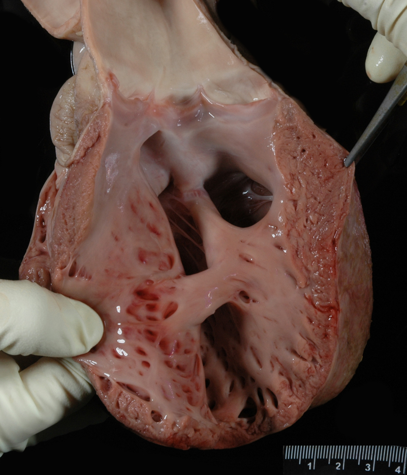
1-8) Many examples of ventricular septal defects!
𝐒𝐨𝐮𝐫𝐜𝐞𝐬
Maxie, G. Jubb, Kennedy and Palmer’s Pathology of Domestic Animals, Volume 1. Sixth Edition.
Photos 1-2 © University of Calgary Diagnostic Services Unit.
Photos 3-8 © Noah’s Arkive contributors Miller, Leininger, Leipold, Morris licensed under CC BY-SA 4.0.


