


Today’s path rounds are on 𝐡𝐲𝐝𝐫𝐨𝐜𝐞𝐩𝐡𝐚𝐥𝐮𝐬!
𝐖𝐡𝐚𝐭 𝐢𝐬 𝐢𝐭?
Hydrocephalus is the abnormal accumulation of fluid in the cranial cavity, in most cases within the brain’s 𝐯𝐞𝐧𝐭𝐫𝐢𝐜𝐥𝐞𝐬 (spaces where the cerebrospinal fluid flows within the brain).
𝐖𝐡𝐨 𝐠𝐞𝐭𝐬 𝐢𝐭?
Any species can get these! Most commonly we see this in dogs, cattle and occasionally in horses.
𝐖𝐡𝐚𝐭 𝐜𝐚𝐮𝐬𝐞𝐬 𝐢𝐭?
There are many different causes of hydrocephalus, but the basic cause is disrupted drainage of cerebrospinal fluid from the brain. This can be caused by a tumour, inflammation, or even a malformation of the brain during development.
𝐖𝐡𝐲 𝐢𝐬 𝐭𝐡𝐢𝐬 𝐚 𝐩𝐫𝐨𝐛𝐥𝐞𝐦?
The end result of disrupted drainage is accumulation of fluid within the brain. This leads to 𝐝𝐢𝐥𝐚𝐭𝐢𝐨𝐧 (enlargement) of the ventricles due to the increased fluid pressure. Because the brain is within an enclosed space, the excess fluid accumulation causes compression of the brain tissue by pushing it against the skull. In a developing animal, this pressure can also lead to thinning and softening of the skull bone, making the head very susceptible to trauma.
𝐇𝐨𝐰 𝐢𝐬 𝐢𝐭 𝐝𝐢𝐚𝐠𝐧𝐨𝐬𝐞𝐝?
In a young animal, the head often has a domed shaped with a softened skull, which can lead to a veterinarian making an initial diagnosis based off clinical suspicion. Animals may have 𝐧𝐞𝐮𝐫𝐨𝐥𝐨𝐠𝐢𝐜 𝐬𝐢𝐠𝐧𝐬, like falling down or extreme lethargy, pointing to issues within the brain. To fully diagnose the condition in a living animal, X-rays or an MRI can be conducted to visualize the excess fluid. At necropsy, the skull cap can be removed to visualize the brain directly. These brains often look soft and floppy, not firm like they normally would. From there, the brain can be removed and sliced into sections to visualize the size of the ventricles, confirming the diagnosis.
𝐇𝐨𝐰 𝐢𝐬 𝐢𝐭 𝐭𝐫𝐞𝐚𝐭𝐞𝐝?
In some cases, drugs can be administered to reduce how much cerebrospinal fluid is produced, hopefully allowing the brain’s drainage systems to keep up. There are some surgical options for improving drainage available as well, depending on the patient.
𝐏𝐡𝐨𝐭𝐨𝐬
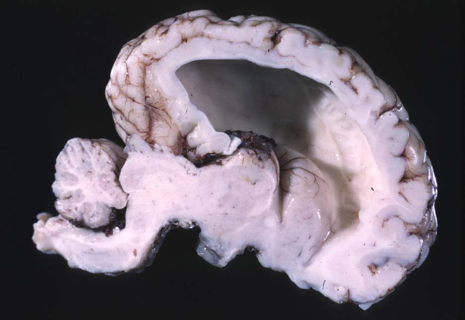
1) The classic appearance of hydrocephalus with a large, empty space within the brain. This photo is just *chef’s kiss*, so beautiful!
2) Another 𝐥𝐨𝐧𝐠𝐢𝐭𝐮𝐝𝐢𝐧𝐚𝐥 𝐬𝐞𝐜𝐭𝐢𝐨𝐧 of the brain showing a dilated ventricle.
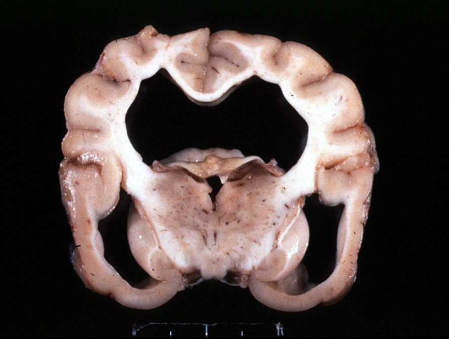
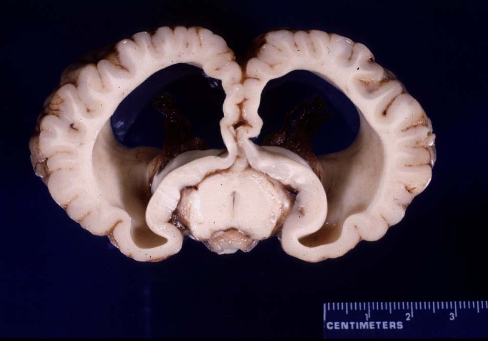
3-4) Two examples of 𝐜𝐫𝐨𝐬𝐬-𝐬𝐞𝐜𝐭𝐢𝐨𝐧𝐬 showing dilated ventricles. Your brain should not have holes that large in it!
5) A fully trimmed normal brain for comparison to the two previous photos.
𝐒𝐨𝐮𝐫𝐜𝐞𝐬
Maxie, G. Jubb, Kennedy and Palmer’s Pathology of Domestic Animals, Volume 2. Sixth Edition.
Photos 1 and 5 courtesy of University of Calgary Diagnostic Services Unit.
Photo 2-4 courtesy of Noah’s Arkive.


