

Today’s path rounds are on 𝐭𝐞𝐭𝐫𝐚𝐥𝐨𝐠𝐲 𝐨𝐟 𝐅𝐚𝐥𝐥𝐨𝐭!
𝐖𝐡𝐚𝐭 𝐢𝐬 𝐢𝐭?
𝐓𝐞𝐭𝐫𝐚𝐥𝐨𝐠𝐲 𝐨𝐟 𝐅𝐚𝐥𝐥𝐨𝐭 is a combination of four 𝐜𝐨𝐧𝐠𝐞𝐧𝐢𝐭𝐚𝐥 (present at birth) defects in the heart, leading to abnormal blood flow. This condition is quite rare, but interesting!
𝐖𝐡𝐨 𝐠𝐞𝐭𝐬 𝐢𝐭?
Any species can get this!
𝐖𝐡𝐚𝐭 𝐜𝐚𝐮𝐬𝐞𝐬 𝐢𝐭?
Tetralogy means 𝐟𝐨𝐮𝐫 𝐰𝐨𝐫𝐤𝐬, and is most often used in literature (i.e. it comes after a trilogy ![]() ). In the case of tetralogy of Fallot, the “four works” are the four different congenital abnormalities present in the disease. These defects are:
). In the case of tetralogy of Fallot, the “four works” are the four different congenital abnormalities present in the disease. These defects are:
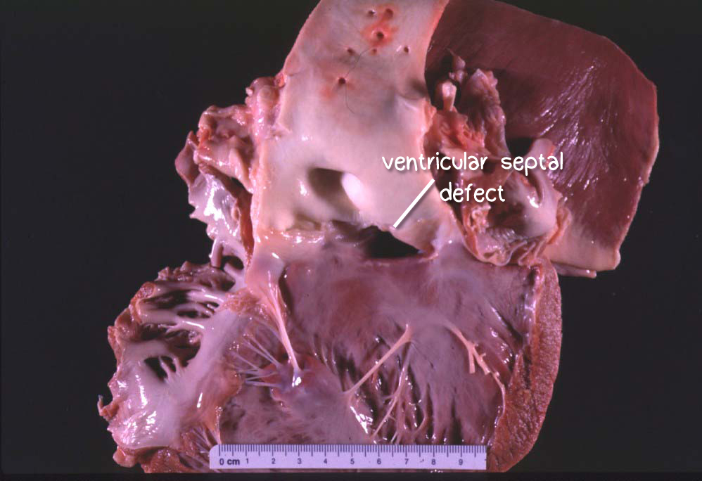
1. 𝐕𝐞𝐧𝐭𝐫𝐢𝐜𝐮𝐥𝐚𝐫 𝐬𝐞𝐩𝐭𝐚𝐥 𝐝𝐞𝐟𝐞𝐜𝐭, a hole in the separation between the two halves of the heart.
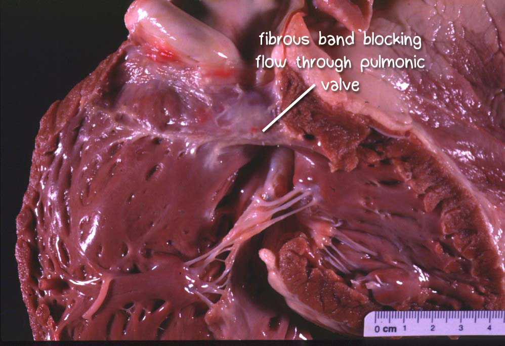
2. 𝐏𝐮𝐥𝐦𝐨𝐧𝐢𝐜 𝐬𝐭𝐞𝐧𝐨𝐬𝐢𝐬, or narrowing of the valve that opens and closes to regulate blood entering the lungs for oxygenation.
3. 𝐎𝐯𝐞𝐫𝐫𝐢𝐝𝐢𝐧𝐠 𝐚𝐨𝐫𝐭𝐚, or an aorta that emerges from both sides of the heart, rather than just the left side.
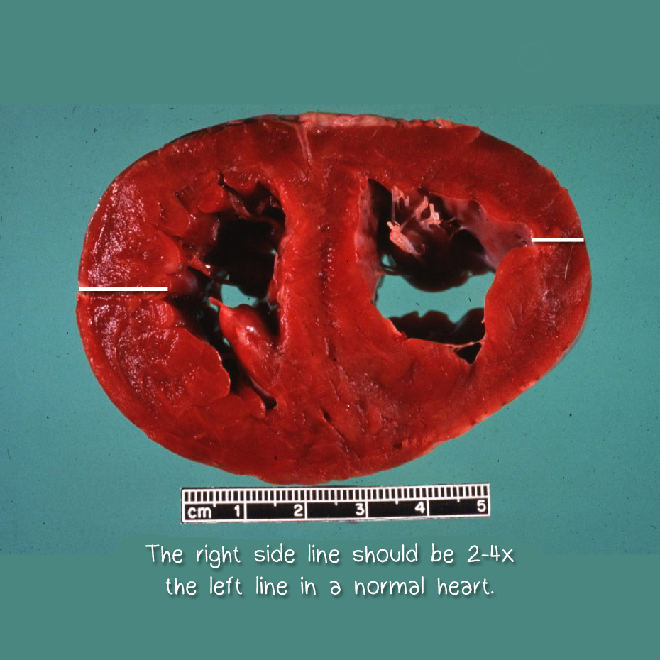
4. 𝐑𝐢𝐠𝐡𝐭 𝐯𝐞𝐧𝐭𝐫𝐢𝐜𝐮𝐥𝐚𝐫 𝐡𝐲𝐩𝐞𝐫𝐭𝐫𝐨𝐩𝐡𝐲, or thickening of the muscle wall on the right side of the heart.
This combination of abnormalities is typically a 𝐬𝐩𝐨𝐫𝐚𝐝𝐢𝐜 (random) occurrence, however it has been found to have a genetic link in Keeshond breed dogs.
𝐖𝐡𝐲 𝐢𝐬 𝐭𝐡𝐢𝐬 𝐚 𝐩𝐫𝐨𝐛𝐥𝐞𝐦?
The main issue with tetralogy of Fallot is 𝐡𝐲𝐩𝐨𝐱𝐢𝐚, or inadequate oxygenation of the blood. The main cause of this is inadequate delivery of blood to the lungs from the pulmonary stenosis, preventing proper oxygenation. This effect is enhanced by the ventricular septal defect, which allows deoxygenated blood to cross into the left side of the heart, mix with oxygenated blood, and be delivered to the body. This poorly oxygenated mixture is not very helpful for the tissues that desperately need oxygen!
In order to try and compensate for the lack of oxygen, the kidneys produce 𝐞𝐫𝐲𝐭𝐡𝐫𝐨𝐩𝐨𝐢𝐞𝐭𝐢𝐧, a hormone that stimulates red blood cell production. Ultimately, this results in 𝐩𝐨𝐥𝐲𝐜𝐲𝐭𝐡𝐞𝐦𝐢𝐚, or having too many red blood cells. The blood becomes viscous and thick, and cannot flow into smaller vessels. This can lead to seizures and 𝐚𝐭𝐚𝐱𝐢𝐚 (irregular gait).
𝐇𝐨𝐰 𝐢𝐬 𝐢𝐭 𝐝𝐢𝐚𝐠𝐧𝐨𝐬𝐞𝐝?
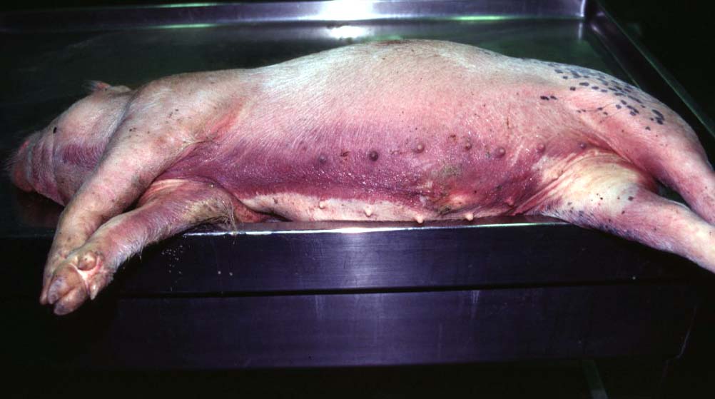
Often, the main clinical sign a veterinarian will see on physical exam is 𝐜𝐲𝐚𝐧𝐨𝐬𝐢𝐬, or blue tissue. This is because the animal doesn’t have enough oxygen in their blood, which makes the blood appear red! These animals also have a history of collapse, seizures, and difficulty breathing. To confirm the diagnosis, 𝐞𝐜𝐡𝐨𝐜𝐚𝐫𝐝𝐢𝐨𝐠𝐫𝐚𝐩𝐡𝐲 (ultrasound of the heart) can be used to identify the abnormalities directly.
𝐇𝐨𝐰 𝐢𝐬 𝐢𝐭 𝐭𝐫𝐞𝐚𝐭𝐞𝐝?
Although there are surgical procedures that can help, this is very rarely done in our animal patients. Sometimes, these animals are managed with medication to improve their quality of life. However, this disease has an extremely poor prognosis, so euthanasia is often the outcome for these patients.
𝐏𝐡𝐨𝐭𝐨𝐬
1-4) Examples of the lesions of tetralogy of Fallot!
5) A pig example of cyanosis.
𝐒𝐨𝐮𝐫𝐜𝐞𝐬
Maxie, G. Jubb, Kennedy and Palmer’s Pathology of Domestic Animals, Volume 3. Sixth Edition.
Tou SP. Tetralogy of Fallot in Animals. Merck Veterinary Manual 2020.
Photos 1-5 © Noah’s Arkive contributors Ramos-Vera, Harrington, Andreasen, Domingo licensed under CC BY-SA 4.0.


