


Today’s path rounds is on 𝐩𝐨𝐥𝐢𝐨𝐞𝐧𝐜𝐞𝐩𝐡𝐚𝐥𝐨𝐦𝐚𝐥𝐚𝐜𝐢𝐚! Inspired by a case that came in this week, wasn’t mine though.
𝐓𝐡𝐢𝐬 𝐰𝐨𝐫𝐝 𝐢𝐬 𝐰𝐚𝐲 𝐭𝐨𝐨 𝐥𝐨𝐧𝐠!
Agreed. We typically shorten this disease to just “polio”, for obvious reasons. But if you break it down into its Latin roots, it’s not too bad!
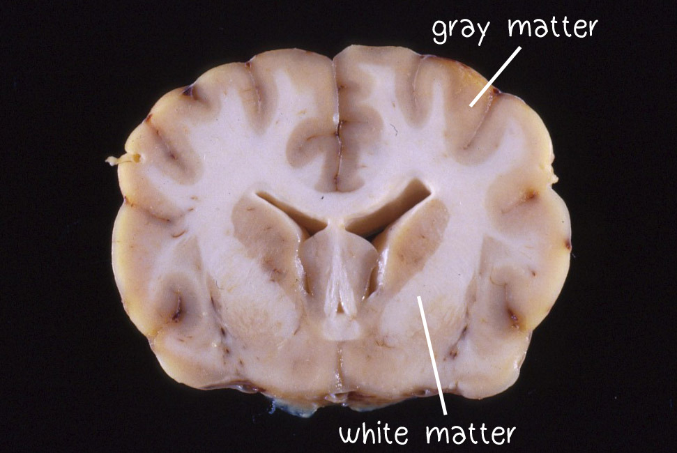
𝐏𝐨𝐥𝐢𝐨 means “gray”, and in this case, refers to the 𝐠𝐫𝐚𝐲 𝐦𝐚𝐭𝐭𝐞𝐫 of the brain. The gray matter is the outer layer of the brain, where all of the 𝐜𝐞𝐥𝐥 𝐛𝐨𝐝𝐢𝐞𝐬 (main processing center) of the neurons live. The center of the brain is the 𝐰𝐡𝐢𝐭𝐞 𝐦𝐚𝐭𝐭𝐞𝐫, which contains all of the 𝐚𝐱𝐨𝐧𝐬 (the main electrical conducting fibre of neurons). So in this disease, the gray matter is primarily affected!
𝐄𝐧𝐜𝐞𝐩𝐡𝐚𝐥𝐨 means brain. This is an important distinction to make, because there is also gray and white matter in the spinal cord. If this disease affected the spinal cord, we would use 𝐦𝐲𝐞𝐥𝐨.
𝐌𝐚𝐥𝐚𝐜𝐢𝐚 means necrosis, which is death of the tissue. So if you put it all together, polioencephalomalacia means death of the gray matter of the brain! Isn’t Latin fun?
𝐖𝐡𝐨 𝐠𝐞𝐭𝐬 𝐢𝐭?
Well, there are a lot of potential causes of this disease across different species, so today we are going to focus on ruminants, i.e. cows, goats, sheep, etc. It most commonly occurs in feedlot animals, however it can also be seen in pastured animals as well.
𝐖𝐡𝐚𝐭 𝐜𝐚𝐮𝐬𝐞𝐬 𝐢𝐭?
The main cause of polio is deficiency of 𝐭𝐡𝐢𝐚𝐦𝐢𝐧𝐞. You might know thiamine as Vitamin B1! Its main function is to help the body process proteins, fats and sugars.
Animals can become thiamine deficient due to low levels in the diet, or perhaps there is something going on that prevents them from absorbing thiamine appropriately. For example, some plant species contain 𝐭𝐡𝐢𝐚𝐦𝐢𝐧𝐚𝐬𝐞, which is an enzyme that destroys thiamine. So animals that are grazing plants with thiaminase will have significantly less thiamine available for their guts to absorb, causing a deficiency.
Similarly, 𝐬𝐮𝐥𝐟𝐮𝐫 can affect thiamine levels in the gut. Sulfur is able to break thiamine in half, causing it to become inactive. Animals ingesting high levels of sulfur through drinking water or feed can also suffer from thiamine deficiency.
𝐇𝐨𝐰 𝐢𝐬 𝐢𝐭 𝐝𝐢𝐚𝐠𝐧𝐨𝐬𝐞𝐝?
Animals with polio will be blind, stop eating, and may press their heads up against walls. As the disease progresses, animals may not be able to walk properly, have muscle tremors and twitching, and eventually may begin seizuring and won’t be able to get up. This disease is typically suspected based on the clinical signs, but a necropsy will be required to confirm the diagnosis.
At necropsy, the pathologist will see areas of the gray matter of the brain that are softer and possibly discoloured yellow. Usually, these areas are roughly symmetrical between the two halves of the brain. The coolest part about polio though, is that the necrotic areas 𝐚𝐮𝐭𝐨𝐟𝐥𝐮𝐨𝐫𝐞𝐬𝐜𝐞, meaning that they glow under UV light! Not all brains with polio will do this, but if you see this change, you can be quite confident about the diagnosis!
𝐏𝐡𝐨𝐭𝐨𝐬
1) A normal brain showing the distinction between the gray and white matter.
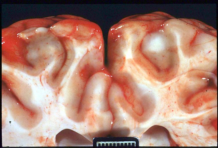
2 and 3) Cross-sections of the brain showing very gooey and flat looking gray matter.
4, 5 and 6) Glowing brains!!!!! Science is so cool. The highlighted regions will correspond with the areas of gooey gray matter.
𝐒𝐨𝐮𝐫𝐜𝐞𝐬
Maxie, G. Jubb, Kennedy and Palmer’s Pathology of Domestic Animals, Volume 1. Sixth Edition.
Photo 4 courtesy of University of Calgary Veterinary Medicine Diagnostic Services Unit.
Photos 1-3, 5 and 6 courtesy of Noah’s Arkive.


