Today’s path rounds is on 𝐧𝐚𝐯𝐢𝐜𝐮𝐥𝐚𝐫 𝐬𝐲𝐧𝐝𝐫𝐨𝐦𝐞/𝐜𝐚𝐮𝐝𝐚𝐥 𝐡𝐞𝐞𝐥 𝐩𝐚𝐢𝐧! This was a request ![]() It’s not something we commonly go digging for in pathology, but it’s still kind of neat!
It’s not something we commonly go digging for in pathology, but it’s still kind of neat!
𝐖𝐡𝐚𝐭’𝐬 𝐚 𝐧𝐚𝐯𝐢𝐜𝐮𝐥𝐚𝐫?
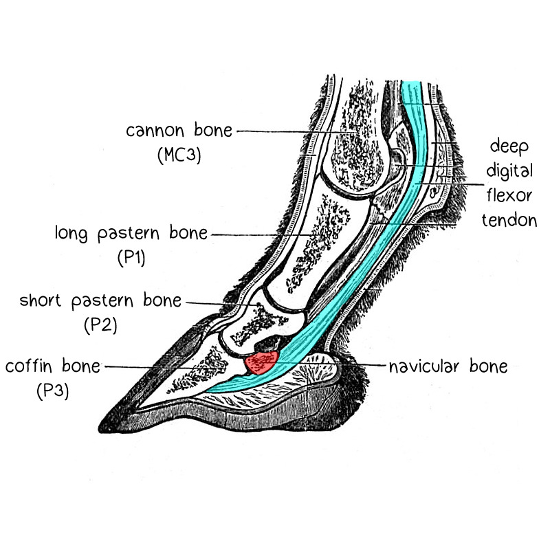
The navicular bone is a tiny little bone in the horse’s foot. Its main function is to help increase the leverage of the 𝐝𝐞𝐞𝐩 𝐝𝐢𝐠𝐢𝐭𝐚𝐥 𝐟𝐥𝐞𝐱𝐨𝐫 𝐭𝐞𝐧𝐝𝐨𝐧 (one of the major flexing tendons that runs down the back of the leg), basically by acting like a pulley to change the direction of the force. Being such a tiny, yet super important bone in such a large horse means it is prone to damage and causing pain when that horse walks.
𝐖𝐡𝐚𝐭’𝐬 𝐧𝐚𝐯𝐢𝐜𝐮𝐥𝐚𝐫 𝐬𝐲𝐧𝐝𝐫𝐨𝐦𝐞?
Navicular syndrome is basically when the navicular bone starts to degenerate. Typically, this means that the cartilage that sits on top of the bone, and sometimes even the bone itself, gets worn down. To try and compensate for the wearing, and desperately try to maintain the normal function of the structure, the body produces extra bone in irregular patches and nodules called 𝐨𝐬𝐭𝐞𝐨𝐩𝐡𝐲𝐭𝐞𝐬. These can help, but sometimes they end up making things worse! The osteophytes can cause damage to the deep digital flexor tendon, leading to scarring that can sometimes even attach the tendon to the bone. Since the tendon is supposed to slide over the bone, this leads to complete loss of function of this area.
𝐖𝐡𝐨 𝐠𝐞𝐭𝐬 𝐢𝐭?
Quarter Horses are the typical patients seen with navicular syndrome, possibly related to their smaller-than-expected hooves in general. However, it is one of the most common causes of forelimb lameness in horses, so pretty much any horse can get it!
𝐖𝐡𝐚𝐭 𝐜𝐚𝐮𝐬𝐞𝐬 𝐢𝐭?
To be honest, no one is really sure. There are three current theories:
1) A blockage of an artery leading to the bone, causing the bone tissue to die.
2) Excessive pressure between the DDFT and the bone due to conformation, hoof size or shoeing technique causing degeneration over time.
3) Process similar to osteoarthritis where there are degenerative changes to the cartilage and bone.
The third theory is the most commonly accepted, but research is still needed to determine a definitive cause!
𝐇𝐨𝐰 𝐢𝐬 𝐢𝐭 𝐝𝐢𝐚𝐠𝐧𝐨𝐬𝐞𝐝?
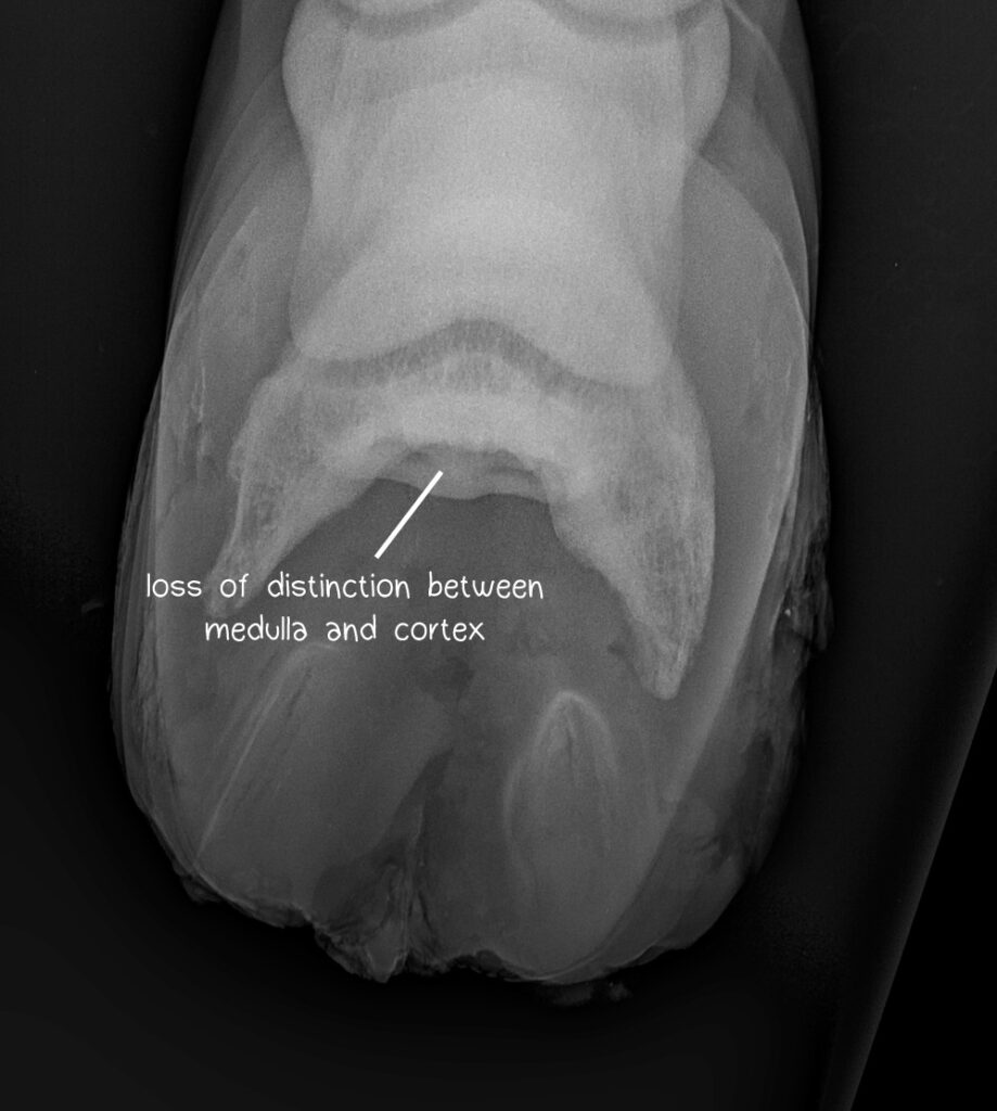
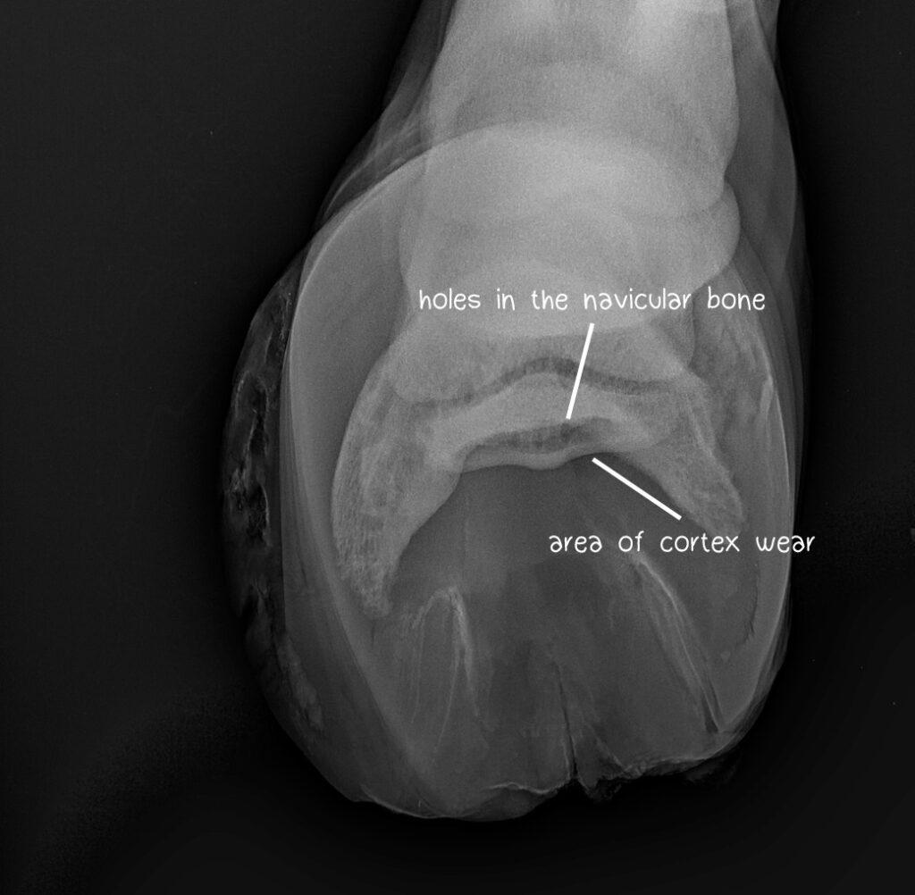
This disease is most commonly diagnosed by a lameness exam followed up by X-rays. On lameness exam, the hallmark sign is lameness that gets significantly worse when the horse is turning towards the painful leg. Sometimes horses with this condition can be pretty much sound on the straightaway! On X-rays, close examination of the navicular bone can show the osteophytes mentioned previously, large hole-like structures, loss of the normal distinct line between the 𝐜𝐨𝐫𝐭𝐞𝐱 (outer section of bone, the hard stuff) and 𝐦𝐞𝐝𝐮𝐥𝐥𝐚 (inner section of bone, where the soft bone marrow is), and sometimes decreased thickness of the cortex due to wear.
𝐖𝐡𝐚𝐭’𝐬 𝐢𝐧 𝐚 𝐧𝐚𝐦𝐞?
An important thing to know about navicular syndrome is that the name can be controversial… in fact, the veterinary community is moving towards calling the suite of clinical signs associated with this condition “caudal heel pain”. Why? Because unlike what the name “navicular syndrome” suggests, there are actually a ton of structures in the foot besides the navicular bone that can cause similar clinical signs. So unless the navicular bone can be specifically identified as the cause of the pain, “CHP” is the most accurate terminology.
𝐏𝐡𝐨𝐭𝐨𝐬
1) A diagram showing the location of the navicular bone relative to the deep digital flexor tendon.
2 and 3) X-rays of a horse with relatively mild navicular syndrome, showing some of the changes that might be seen.
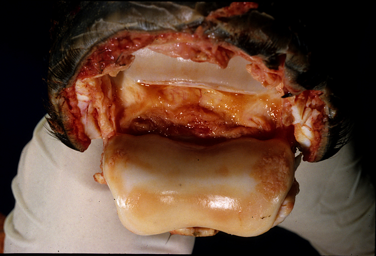
4) The surface of a navicular bone showing cartilage wear down to the underlying bone on the outer edges (the areas that look very rough!).
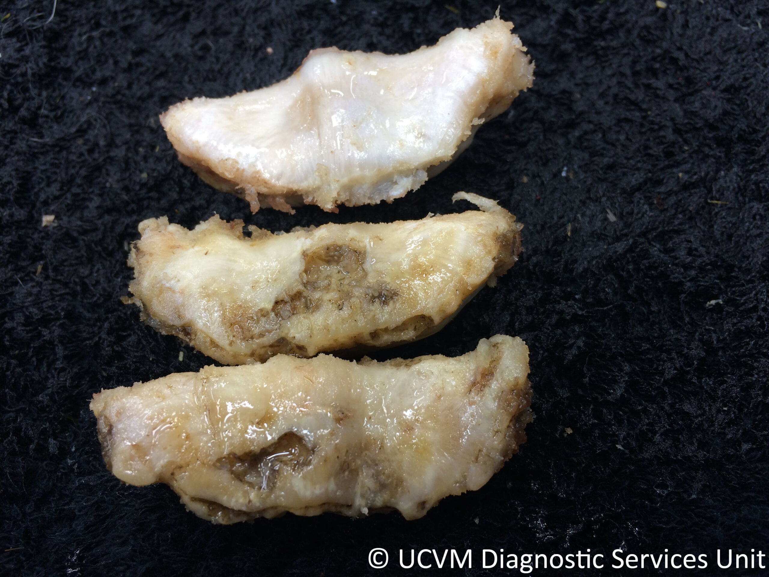
5) Some really garbage navicular bones, with a normal comparison at the top.
𝐒𝐨𝐮𝐫𝐜𝐞𝐬
Maxie, G. Jubb, Kennedy and Palmer’s Pathology of Domestic Animals, Volume 1. Sixth Edition.
Photo 4 courtesy of Noah’s Arkive.
Photo 5 courtesy of University of Calgary Veterinary Medicine Diagnostic Services Unit.


