Today’s path rounds are on 𝐟𝐞𝐥𝐢𝐧𝐞 𝐜𝐨𝐫𝐧𝐞𝐚𝐥 𝐬𝐞𝐪𝐮𝐞𝐬𝐭𝐫𝐚! This was a request ![]()
𝐖𝐡𝐚𝐭 𝐢𝐬 𝐢𝐭?
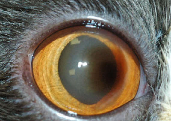
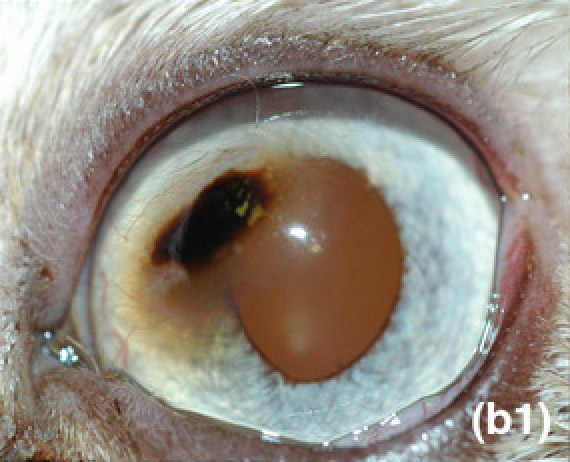
𝐅𝐞𝐥𝐢𝐧𝐞 𝐜𝐨𝐫𝐧𝐞𝐚𝐥 𝐬𝐞𝐪𝐮𝐞𝐬𝐭𝐫𝐚 are an orange-brown area of 𝐧𝐞𝐜𝐫𝐨𝐬𝐢𝐬 (cell death) on the 𝐜𝐨𝐫𝐧𝐞𝐚 (outer, clear layer of the eye). They can be 𝐰𝐞𝐥𝐥-𝐝𝐞𝐦𝐚𝐫𝐜𝐚𝐭𝐞𝐝 (well defined borders) or more “fuzzy” looking. Typically only one eye is affected, however both eyes can be affected either at the same time or at different times.
𝐖𝐡𝐨 𝐠𝐞𝐭𝐬 𝐢𝐭?
This condition is seen in cats, particularly Persian and Himalayan cats.
𝐖𝐡𝐚𝐭 𝐜𝐚𝐮𝐬𝐞𝐬 𝐢𝐭?
The exact cause of these sequestra is unknown, but it seems likely that it is a 𝐬𝐞𝐪𝐮𝐞𝐥 (condition arising from another condition) of corneal ulceration, which is basically a large hole in the cornea. This can be caused by chronic dry eye, viral diseases, or trauma.
𝐖𝐡𝐲 𝐢𝐬 𝐭𝐡𝐢𝐬 𝐚 𝐩𝐫𝐨𝐛𝐥𝐞𝐦?
These lesions are incredibly painful for the cat, and in some cases can even lead to 𝐠𝐥𝐨𝐛𝐞 𝐫𝐮𝐩𝐭𝐮𝐫𝐞 (rupture of the eyeball ![]() ) if the lesion penetrates all the way through the cornea.
) if the lesion penetrates all the way through the cornea.
𝐖𝐡𝐲 𝐢𝐬 𝐢𝐭 𝐜𝐨𝐥𝐨𝐮𝐫𝐟𝐮𝐥?
The pigment that makes the lesion orangey to black are actually from 𝐩𝐨𝐫𝐩𝐡𝐲𝐫𝐢𝐧𝐬 (organic compounds) that are found within the normal tear film that covers the eye. Because there’s a defect in the corneal tissue, the porphyrins accumulate there, making an orange to black colour!
𝐇𝐨𝐰 𝐢𝐬 𝐢𝐭 𝐝𝐢𝐚𝐠𝐧𝐨𝐬𝐞𝐝?
These lesions are pretty easy to see, and there isn’t much else that looks like them! To help confirm the diagnosis, the veterinarian can do a complete ophthalmic exam to assess the whole eye.
𝐇𝐨𝐰 𝐢𝐬 𝐢𝐭 𝐭𝐫𝐞𝐚𝐭𝐞𝐝?
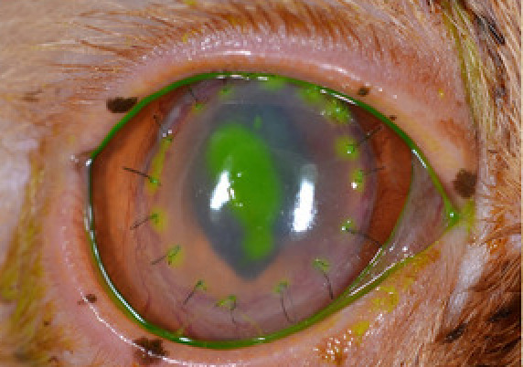
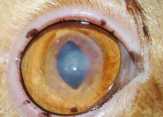
These lesions are typically treated by 𝐤𝐞𝐫𝐚𝐭𝐞𝐜𝐭𝐨𝐦𝐲, or removal of a layer of the cornea. After that, the hole in the cornea is usually patched with some sort of graft. Veterinarians have successfully used 𝐜𝐨𝐧𝐣𝐮𝐧𝐜𝐭𝐢𝐯𝐚 (the tissue around the eye), porcine corneal tissue and even porcine intestine to make these grafts. Crazy!
𝐏𝐡𝐨𝐭𝐨𝐬
1-5) Examples of corneal sequestra ranging from well-demarcated to more amorphous.
6-8) A series of photos showing a lesion (the bright green stuff is fluorescent dye used to identify ulcers), the lesion after receiving a graft from a donor eye, and finally the healed lesion!
𝐒𝐨𝐮𝐫𝐜𝐞𝐬
Laguna, F., Leiva, M., Costa, D., Lacerda, R., Gimenez, T.P. Corneal grafting for the treatment of feline corneal sequestrum: a retrospective study of 18 eyes (13 cats). Veterinary Ophthalmology (2015) 18, 4, 291-296.
Featherstone, H.J., Sansom, J. Feline corneal sequestra: a review of 64 cases (80 eyes) from 1993 to 2000. Veterinary Ophthalmology (2004) 7, 4, 213-227.
Andrew, S.E., Tou, S., Brooks, D.E. Corneoconjunctival transposition for the treatment of feline corneal sequestra: a retrospective study of 17 cases (1990-1998). Veterinary Ophthalmology (2001) 4, 2, 107-111.
Photo 1 from Andrew et al. Photo 2-4 from Featherstone et al.
Photos 5-8 from Laguna et al.
