Today’s path rounds are on 𝐞𝐧𝐳𝐨𝐨𝐭𝐢𝐜 𝐧𝐚𝐬𝐚𝐥 𝐭𝐮𝐦𝐨𝐮𝐫! This was a request ![]()
𝐖𝐡𝐚𝐭 𝐢𝐬 𝐢𝐭?
𝐄𝐧𝐳𝐨𝐨𝐭𝐢𝐜 𝐧𝐚𝐬𝐚𝐥 𝐭𝐮𝐦𝐨𝐮𝐫 is a form of 𝐧𝐚𝐬𝐚𝐥 𝐚𝐝𝐞𝐧𝐨𝐜𝐚𝐫𝐜𝐢𝐧𝐨𝐦𝐚 (tumour of the glandular tissue of the nose) that is caused by a viral infection.
𝐖𝐡𝐨 𝐠𝐞𝐭𝐬 𝐢𝐭?
This disease is seen in sheep and goats!
𝐖𝐡𝐚𝐭 𝐜𝐚𝐮𝐬𝐞𝐬 𝐢𝐭?
This tumour is caused by 𝐞𝐧𝐳𝐨𝐨𝐭𝐢𝐜 𝐧𝐚𝐬𝐚𝐥 𝐭𝐮𝐦𝐨𝐮𝐫 𝐯𝐢𝐫𝐮𝐬, which is a retrovirus in the same family as things like HIV. The virus is acquired by contact with nasal secretions from affected animals, and 𝐢𝐧𝐜𝐮𝐛𝐚𝐭𝐞𝐬 (remains dormant) for 1-3 years before developing nasal tumours. That said, it is far more common to have 𝐬𝐮𝐛𝐜𝐥𝐢𝐧𝐢𝐜𝐚𝐥 (not apparent) infection where no tumours are produced, so finding the source of the virus can be very difficult!
𝐖𝐡𝐲 𝐢𝐬 𝐭𝐡𝐢𝐬 𝐚 𝐩𝐫𝐨𝐛𝐥𝐞𝐦?
The tumours arise within the nasal cavity and cause progressive respiratory issues, nasal deformity and nasal discharge. In some cases, the tumour may take up an entire nasal passage and even poke out of the nostril! Particularly aggressive tumours may also invade into the bone and cause deviation of the nasal septum and other boney changes.
𝐇𝐨𝐰 𝐢𝐬 𝐢𝐭 𝐝𝐢𝐚𝐠𝐧𝐨𝐬𝐞𝐝?
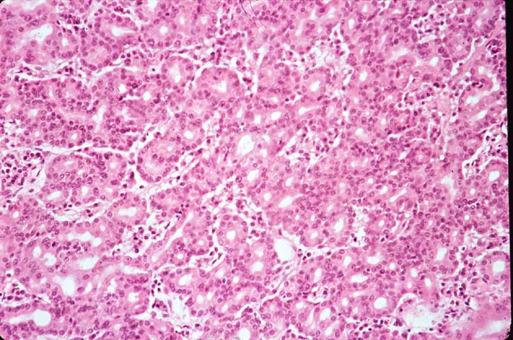
Typically these lesions will be diagnosed based on a 𝐛𝐢𝐨𝐩𝐬𝐲 (sample of tissue) that is sent to a pathologist. Under the microscope, the pathologist can see tubular formations that indicate the tumour cells originated from glandular structures. Super cool! To confirm viral involvement, 𝐏𝐂𝐑 testing to directly identify the virus’ proteins can be used.
𝐇𝐨𝐰 𝐢𝐬 𝐢𝐭 𝐭𝐫𝐞𝐚𝐭𝐞𝐝?
Unfortunately the location of these tumours makes it extremely difficult to remove them surgically. Therefore, these animals are often euthanized for welfare reasons.
𝐏𝐡𝐨𝐭𝐨𝐬
1-4) Examples of nasal tumours taking up space in the nose.
5-6) Lovely examples of the tubular structures that a pathologist would see on histology! Remember we are looking at a 2D section of a 3D structure, so tubes look like circles in our world.
𝐒𝐨𝐮𝐫𝐜𝐞𝐬
Maxie, G. Jubb, Kennedy and Palmer’s Pathology of Domestic Animals, Volume 2. Sixth Edition.
Photos 1-6 courtesy of Noah’s Arkive.


