


Today’s path rounds are on 𝐜𝐡𝐨𝐥𝐚𝐧𝐠𝐢𝐨𝐜𝐚𝐫𝐜𝐢𝐧𝐨𝐦𝐚𝐬!
𝐖𝐡𝐚𝐭 𝐢𝐬 𝐢𝐭?
𝐂𝐡𝐨𝐥𝐚𝐧𝐠𝐢𝐨𝐜𝐚𝐫𝐜𝐢𝐧𝐨𝐦𝐚𝐬 are malignant tumours of the 𝐛𝐢𝐥𝐢𝐚𝐫𝐲 𝐞𝐩𝐢𝐭𝐡𝐞𝐥𝐢𝐮𝐦 (the cells that line the bile ducts in the liver).
𝐖𝐡𝐨 𝐠𝐞𝐭𝐬 𝐢𝐭?
Any species can get these! Most commonly they are reported in dogs, cats, sheep, cattle, horses and goats.
𝐖𝐡𝐲 𝐢𝐬 𝐭𝐡𝐢𝐬 𝐚 𝐩𝐫𝐨𝐛𝐥𝐞𝐦?
These tumours can often take up a large amount of the liver, causing damage. This leads to 𝐥𝐢𝐯𝐞𝐫 𝐟𝐚𝐢𝐥𝐮𝐫𝐞, which can lead to all kinds of downstream effects that are too numerous for this post. Additionally, these tumours commonly metastasize to nearby lymph nodes, the lungs and the 𝐩𝐞𝐫𝐢𝐭𝐨𝐧𝐞𝐮𝐦 (the thin membrane that helps keep the intestines organized).
𝐇𝐨𝐰 𝐢𝐬 𝐢𝐭 𝐝𝐢𝐚𝐠𝐧𝐨𝐬𝐞𝐝?
Typically these animals will present to their veterinarian with 𝐣𝐚𝐮𝐧𝐝𝐢𝐜𝐞, or yellowing of the tissues due to accumulation of the yellow pigment 𝐛𝐢𝐥𝐢𝐫𝐮𝐛𝐢𝐧. Bilirubin is normally processed by the liver and excreted in the bile, so if the liver is damaged, then bilirubin accumulates in the tissues causing them to turn yellow. Based on this finding on physical exam and bloodwork showing liver issues, the veterinarian may suspect that there is some type of liver disease, leading to an ultrasound. On ultrasound, the liver masses can be visualized and sampled for submission to a pathologist to confirm the diagnosis.
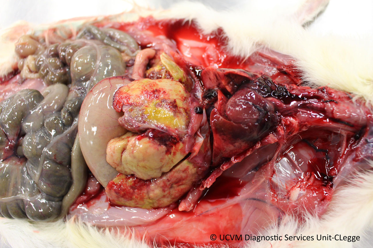
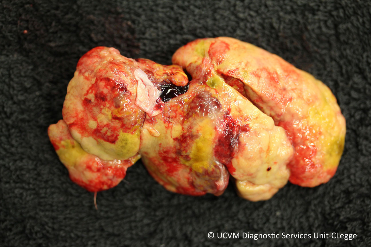
At necropsy, cholangiocarcinomas tend to have an 𝐮𝐦𝐛𝐢𝐥𝐢𝐜𝐚𝐭𝐞𝐝 appearance, meaning they have a raised rim of tissue with a central depression, like a belly button! Based on this appearance, the pathologist can make an initial diagnosis, and then later confirm it on 𝐡𝐢𝐬𝐭𝐨𝐥𝐨𝐠𝐲 (looking at the tissue under a microscope).
𝐇𝐨𝐰 𝐢𝐬 𝐢𝐭 𝐭𝐫𝐞𝐚𝐭𝐞𝐝?
Depending on the severity of disease, the masses may be able to be removed surgically. Unfortunately, even despite surgical treatment, the prognosis for these patients is very poor.
𝐏𝐡𝐨𝐭𝐨𝐬
1) The classic appearance of umbilicated cholangiocarcinoma.
2-3) A very severe case of cholangiocarcinoma in a guinea pig.
4-5) A couple more examples!
𝐒𝐨𝐮𝐫𝐜𝐞𝐬
Maxie, G. Jubb, Kennedy and Palmer’s Pathology of Domestic Animals, Volume 2. Sixth Edition.
Photos 1-3 courtesy of University of Calgary Diagnostic Services Unit.
Photo 4-5 courtesy of Noah’s Arkive.


