Today’s path rounds are on 𝐟𝐢𝐥𝐚𝐫𝐢𝐚𝐥 𝐝𝐞𝐫𝐦𝐚𝐭𝐢𝐭𝐢𝐬!
𝐖𝐡𝐚𝐭 𝐢𝐬 𝐢𝐭?
𝐅𝐢𝐥𝐚𝐫𝐢𝐚𝐥 𝐝𝐞𝐫𝐦𝐚𝐭𝐢𝐭𝐢𝐬 is 𝐝𝐞𝐫𝐦𝐚𝐭𝐢𝐭𝐢𝐬 (inflammation of the skin) caused by 𝐟𝐢𝐥𝐚𝐫𝐢𝐚, which are small worm-like parasites. 𝐒𝐭𝐞𝐩𝐡𝐚𝐧𝐨𝐟𝐢𝐥𝐚𝐫𝐢𝐚 are the most common parasites we see causing this condition!
𝐖𝐡𝐨 𝐠𝐞𝐭𝐬 𝐢𝐭?
This disease occurs in cattle, all over the world!
𝐖𝐡𝐚𝐭 𝐜𝐚𝐮𝐬𝐞𝐬 𝐢𝐭?
As mentioned, this condition is caused by Stephanofilaria parasites. Depending on where you are in the world, different species of this genus affect cattle. In North America, cattle most commonly get 𝐒𝐭𝐞𝐩𝐡𝐚𝐧𝐨𝐟𝐢𝐥𝐚𝐫𝐢𝐚 𝐬𝐭𝐢𝐥𝐞𝐬𝐢.
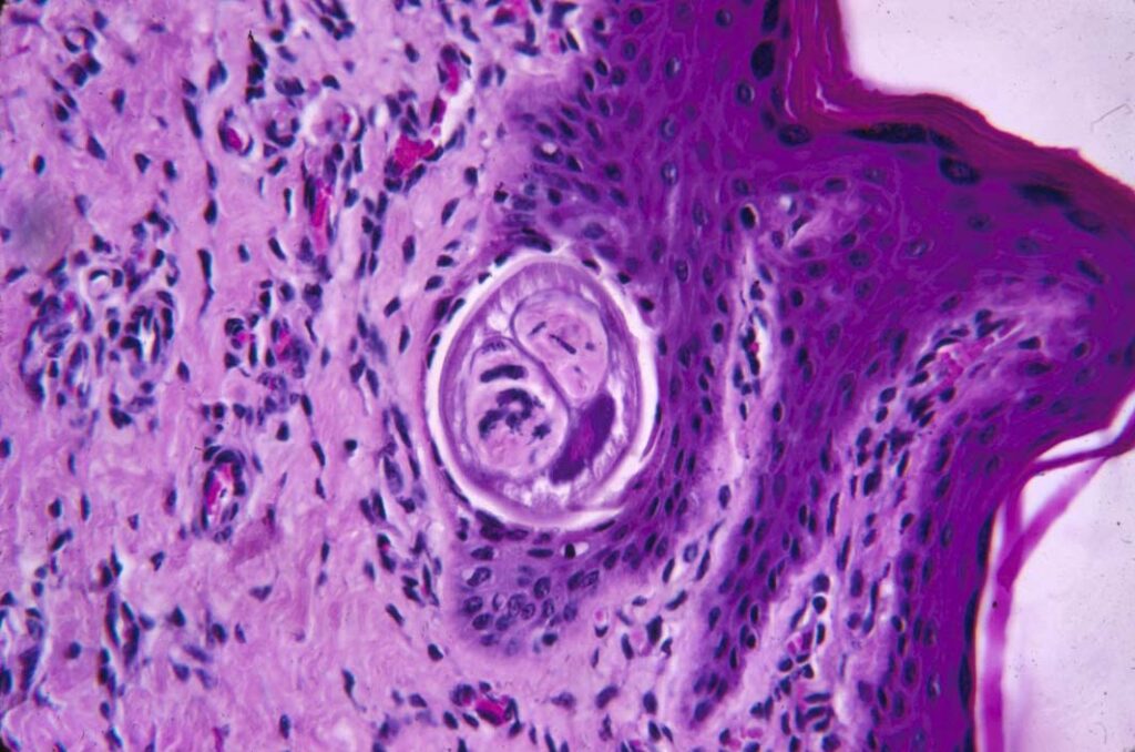
These parasites are transmitted by the 𝐡𝐨𝐫𝐧 𝐟𝐥𝐲, which become infected with the parasite after feeding on an infected cow’s skin wound. The fly picks up 𝐦𝐢𝐜𝐫𝐨𝐟𝐢𝐥𝐚𝐫𝐢𝐚𝐞, a microscopic worm-like stage, which then develop into larvae within the fly. The larvae are deposited onto the skin of an uninfected cow, where they migrate to the hair follicles. Here, they make themselves a home, develop into adults, and begin producing new microfilariae, allowing the process to continue.
𝐖𝐡𝐲 𝐢𝐬 𝐭𝐡𝐢𝐬 𝐚 𝐩𝐫𝐨𝐛𝐥𝐞𝐦?
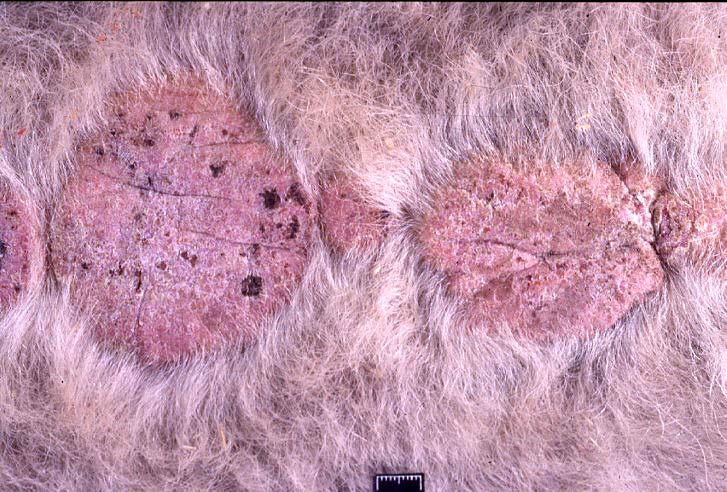
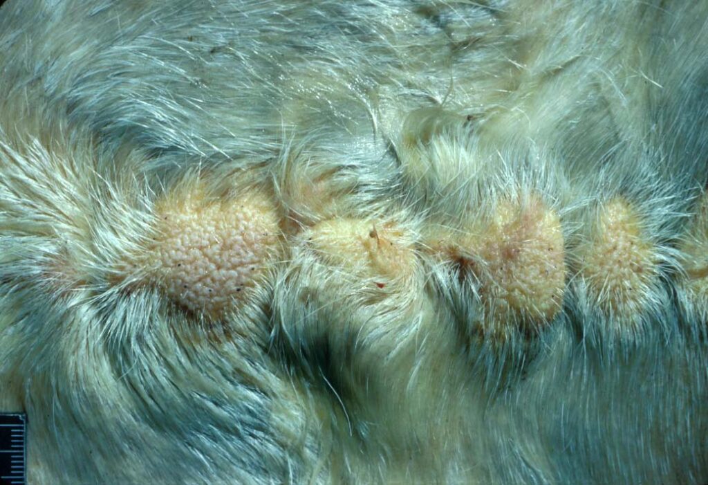
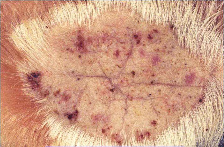
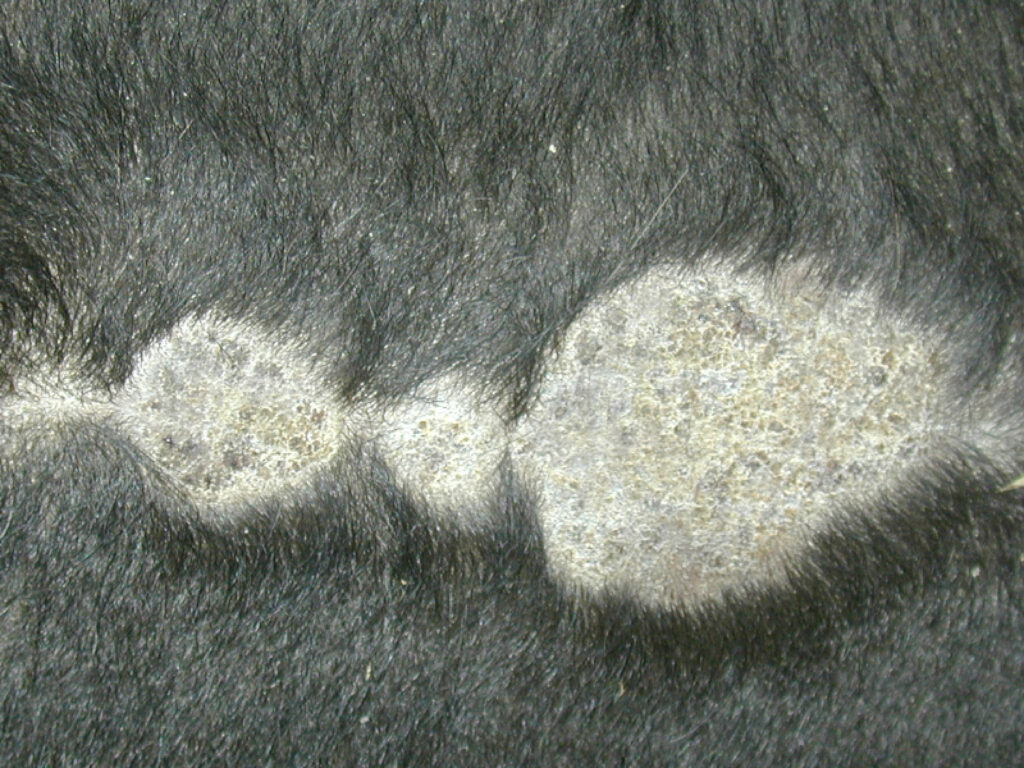
Infection of the hair follicles with these parasites produces circular, hairless scabs or crusts on the animal. Typically we see these on the 𝐯𝐞𝐧𝐭𝐫𝐚𝐥 𝐦𝐢𝐝𝐥𝐢𝐧𝐞 (very middle of the belly) in North America. These lesions typically don’t cause too many issues for the cow, other than they might be a bit itchy. However, they can be an issue for producers who want to sell hides, as they make the hide thickened, rough and generally not very pleasant to look at!
𝐇𝐨𝐰 𝐢𝐬 𝐢𝐭 𝐝𝐢𝐚𝐠𝐧𝐨𝐬𝐞𝐝?
These parasites can be diagnosed by 𝐝𝐞𝐞𝐩 𝐬𝐤𝐢𝐧 𝐬𝐜𝐫𝐚𝐩𝐢𝐧𝐠, where a sharp blade is used to scrape the skin surface and gather cells to look at under the microscope. Under the microscope, the adults or microfilariae can be seen!
𝐇𝐨𝐰 𝐢𝐬 𝐢𝐭 𝐭𝐫𝐞𝐚𝐭𝐞𝐝?
There is no specific treatment for this parasite, but it does seem susceptible to most of our anti-parasite medications typically used in cattle.
𝐏𝐡𝐨𝐭𝐨𝐬
1-4) Examples of the round, crusty, hairless lesions on the belly of cattle.
5) A little friend hanging out in a hair follicle!
𝐒𝐨𝐮𝐫𝐜𝐞𝐬
Maxie, G. Jubb, Kennedy and Palmer’s Pathology of Domestic Animals, Volume 1. Sixth Edition.
Gerhold RW. Stephanofilariasis in Animals. Merck Veterinary Manual 2020.
Photos 1-5 © Noah’s Arkive contributors Read, Vet Diag Lab, Chapman, Williams, McGavin licensed under CC BY-SA 4.0.


