Today’s path rounds are on 𝐰𝐡𝐢𝐭𝐞 𝐦𝐮𝐬𝐜𝐥𝐞 𝐝𝐢𝐬𝐞𝐚𝐬𝐞!
𝐖𝐡𝐚𝐭 𝐢𝐬 𝐢𝐭?
𝐖𝐡𝐢𝐭𝐞 𝐦𝐮𝐬𝐜𝐥𝐞 𝐝𝐢𝐬𝐞𝐚𝐬𝐞 is basically as the name suggests, a disease of the muscle that turns it white instead of its normal red colour. This disease is associated with nutritional problems!
𝐖𝐡𝐨 𝐠𝐞𝐭𝐬 𝐢𝐭?
We most commonly see this disease in young animals like calves, lambs and foals, however any species can get this.
𝐖𝐡𝐚𝐭 𝐜𝐚𝐮𝐬𝐞𝐬 𝐢𝐭?
White muscle disease can be caused by two nutritional issues: 𝐬𝐞𝐥𝐞𝐧𝐢𝐮𝐦 𝐝𝐞𝐟𝐢𝐜𝐢𝐞𝐧𝐜𝐲 or 𝐯𝐢𝐭𝐚𝐦𝐢𝐧 𝐄 𝐝𝐞𝐟𝐢𝐜𝐢𝐞𝐧𝐜𝐲. In mammals, we most commonly associate this condition with selenium deficiency. Both of these nutrients are 𝐚𝐧𝐭𝐢𝐨𝐱𝐢𝐝𝐚𝐧𝐭𝐬, meaning they bind to and neutralize dangerous oxygen-derived chemicals that can damage tissues. When these nutrients are inadequate, the 𝐟𝐫𝐞𝐞 𝐫𝐚𝐝𝐢𝐜𝐚𝐥𝐬 cause damage to the muscular cell membranes, leading to 𝐧𝐞𝐜𝐫𝐨𝐬𝐢𝐬 (cell death). Necrotic tissues commonly undergo 𝐦𝐢𝐧𝐞𝐫𝐚𝐥𝐢𝐳𝐚𝐭𝐢𝐨𝐧, where calcium is deposited in the area, which leads to the muscle turning white!
𝐖𝐡𝐲 𝐢𝐬 𝐭𝐡𝐢𝐬 𝐚 𝐩𝐫𝐨𝐛𝐥𝐞𝐦?
Obviously, muscle that is dead or dying doesn’t function as effectively as a normal, healthy muscle. Because this disease can affect any muscle in the body, the presenting symptoms can be quite variable! Some animals have difficulty eating due to weakening of the muscles in the tongue, while others may have lameness or strange movement due to weakness of the limb muscles. The most severe presentation comes from necrosis of the heart muscle, leading to heart failure.
𝐇𝐨𝐰 𝐢𝐬 𝐢𝐭 𝐝𝐢𝐚𝐠𝐧𝐨𝐬𝐞𝐝?
Diagnosis in the live animal usually involves measuring the concentration of selenium or vitamin E in the blood. However, the easiest method of diagnosis is seeing the white, calcified muscle from a necropsy specimen under the microscope!
𝐇𝐨𝐰 𝐢𝐬 𝐢𝐭 𝐭𝐫𝐞𝐚𝐭𝐞𝐝? 𝐇𝐨𝐰 𝐢𝐬 𝐢𝐭 𝐩𝐫𝐞𝐯𝐞𝐧𝐭𝐞𝐝?
Treatment and prevention of this disease are the same: injecting selenium and vitamin E. In affected animals, this restores the antioxidant capacity, and allows the muscle to begin healing. As a preventative, producers with known issues may give all of their young animals this injection to boost their antioxidants before it becomes a problem!
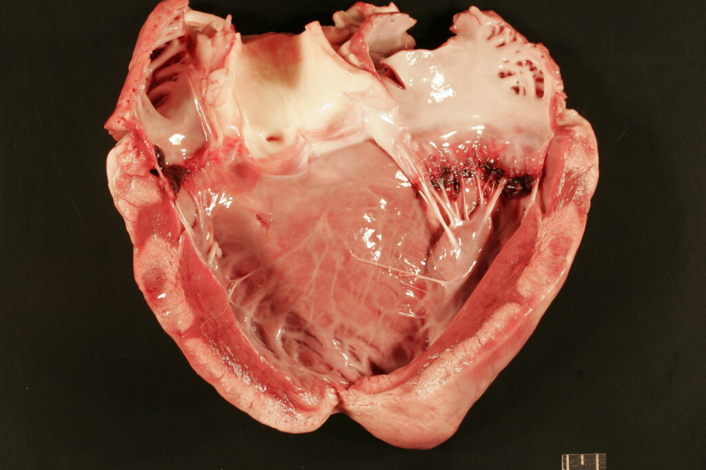
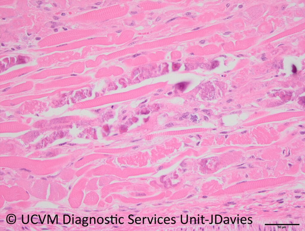
𝐏𝐡𝐨𝐭𝐨𝐬
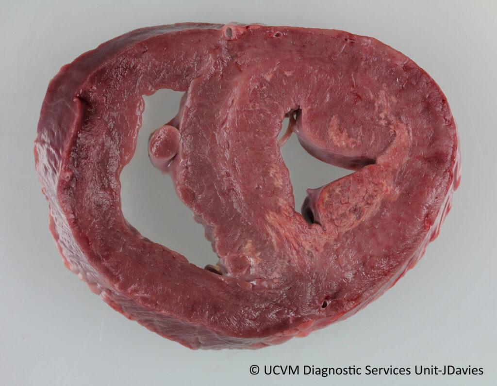
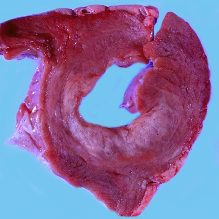
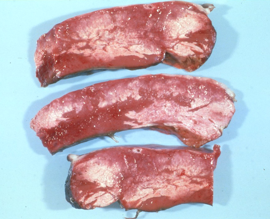
1-4) White muscle in the heart from selenium deficiency.
5) What this disease looks like under the microscope! The normal muscle is bright pink with striping. In the middle, you can see muscle with purple calcium covering it.
𝐒𝐨𝐮𝐫𝐜𝐞𝐬
Maxie, G. Jubb, Kennedy and Palmer’s Pathology of Domestic Animals, Volume 1. Sixth Edition.
Photos 1, 5 © University of Calgary Diagnostic Services Unit.
Photos 2-4 Noah’s Arkive contributors Rimoldi, Luginbuhl, Anderson licensed under CC BY-SA 4.0.


