

Today’s path rounds are on 𝐭𝐨𝐱𝐨𝐩𝐥𝐚𝐬𝐦𝐨𝐬𝐢𝐬!
𝐖𝐡𝐚𝐭 𝐢𝐬 𝐢𝐭?
𝐓𝐨𝐱𝐨𝐩𝐥𝐚𝐬𝐦𝐚 is a protozoan parasite that we most commonly associate with infectious abortion, however it can also cause 𝐬𝐲𝐬𝐭𝐞𝐦𝐢𝐜 (whole body) disease with serious consequences!
𝐖𝐡𝐨 𝐠𝐞𝐭𝐬 𝐢𝐭?
We most commonly associate this disease with cats, because they are the 𝐝𝐞𝐟𝐢𝐧𝐢𝐭𝐢𝐯𝐞 𝐡𝐨𝐬𝐭, meaning they are the primary species where this protozoa likes to live. However, other species, including humans, can become infected with the parasite, causing disease.
𝐖𝐡𝐚𝐭 𝐜𝐚𝐮𝐬𝐞𝐬 𝐢𝐭?
As mentioned, cats are the primary source of these protozoans. Cats become infected by ingesting 𝐨𝐨𝐜𝐲𝐬𝐭𝐬 (egg forms) from the environment, or eating 𝐛𝐫𝐚𝐝𝐲𝐳𝐨𝐢𝐭𝐞𝐬, the form found within 𝐢𝐧𝐭𝐞𝐫𝐦𝐞𝐝𝐢𝐚𝐭𝐞 𝐡𝐨𝐬𝐭𝐬 like rodents. These intermediate hosts become infected by eating the oocysts. Regardless of the source, this infection allows the protozoan to develop into adult forms in the gut of the infected cat, which eventually produce oocysts that are shed in the feces and contaminate the environment. Similar to the intermediate hosts, these oocysts can then be ingested by non-target species, like humans, birds, herbivores and carnivores, leading to infection.
𝐖𝐡𝐲 𝐢𝐬 𝐭𝐡𝐢𝐬 𝐚 𝐩𝐫𝐨𝐛𝐥𝐞𝐦?
Under most circumstances, the immune system will quickly activate to eliminate the protozoan, preventing disease. However, in cases of 𝐢𝐦𝐦𝐮𝐧𝐨𝐜𝐨𝐦𝐩𝐫𝐨𝐦𝐢𝐬𝐞 (weak immune system), the protozoan may spread to multiple organs via the bloodstream. The 𝐭𝐚𝐜𝐡𝐲𝐳𝐨𝐢𝐭𝐞 stage of the parasite invades the cells, where it forms a big storage vessel for itself and begins to replicate. By living within the cells, the parasite is hidden from the immune system. The parasite can live quite happily in this cyst stage for moths or years. However, as the parasite replicates, eventually the storage vessel becomes too large for the cell, rupturing the cell. This alerts the immune system to the parasite’s presence, and stimulates an immune response that can cause cell death in the area.
Because this organism can be spread to many different organisms, this cell death can lead to many different outcomes. For example, infection of the brain can cause incoordination, tremors, seizures and even 𝐩𝐚𝐫𝐞𝐬𝐢𝐬 (inability to move). If it infects the lungs, it can cause difficulty breathing and pneumonia.
𝐇𝐨𝐰 𝐢𝐬 𝐢𝐭 𝐝𝐢𝐚𝐠𝐧𝐨𝐬𝐞𝐝?
Unfortunately, the clinical signs of this disease aren’t very specific, so additional testing is required to confirm the diagnosis. In live animals, usually the diagnosis is made by 𝐬𝐞𝐫𝐨𝐥𝐨𝐠𝐢𝐜 𝐭𝐞𝐬𝐭𝐢𝐧𝐠, where the blood is tested to see if there are antibodies against the parasite. At necropsy, we can see the cysts formed by the parasites directly, making diagnosis somewhat more simple!
𝐇𝐨𝐰 𝐢𝐬 𝐢𝐭 𝐭𝐫𝐞𝐚𝐭𝐞𝐝?
Treatment typically involves 𝐚𝐧𝐭𝐢-𝐜𝐨𝐜𝐜𝐢𝐝𝐢𝐚𝐥 𝐝𝐫𝐮𝐠𝐬 which target this type of parasite specifically. These drugs can also reduce the number of oocysts produced by cats as a potential preventative measure.
𝐖𝐡𝐚𝐭’𝐬 𝐭𝐡𝐞 𝐝𝐞𝐚𝐥 𝐰𝐢𝐭𝐡 𝐩𝐫𝐞𝐠𝐧𝐚𝐧𝐭 𝐰𝐨𝐦𝐞𝐧?
As mentioned, this parasite can infect humans, and can cause abortion. This is the big concern with pregnant women cleaning litter boxes, as they may become exposed to the oocysts in their cat’s feces, leading to abortion. Pregnant women may also be exposed by interacting with soil, raw meat or unwashed vegetables. Thorough cleaning, hand washing and avoiding these circumstances where possible are the best methods of prevention!
𝐏𝐡𝐨𝐭𝐨𝐬
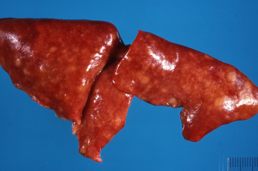
1-2) Pneumonia in a cat with Toxoplasma.
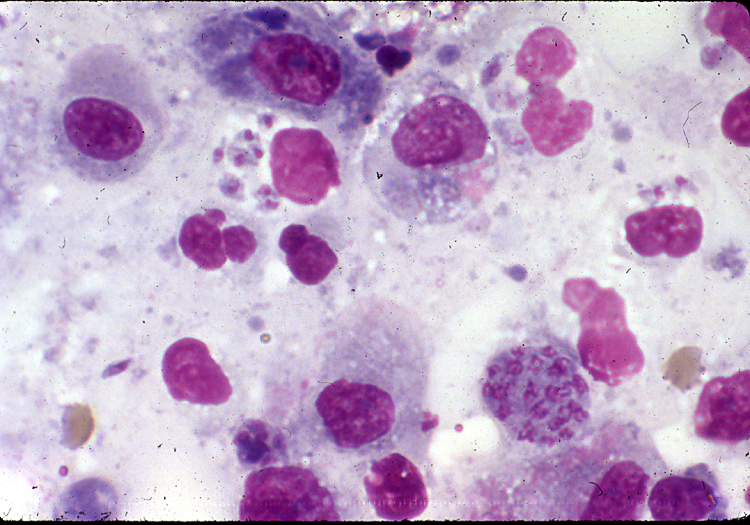
3) Toxoplasma as seen on 𝐜𝐲𝐭𝐨𝐥𝐨𝐠𝐲, where cells are sampled from a live animal.
4) Toxoplasma at 𝐡𝐢𝐬𝐭𝐨𝐥𝐨𝐠𝐲, where the tissue is examined under the microscope! This cyst happens to be in the brain.
𝐒𝐨𝐮𝐫𝐜𝐞𝐬
Maxie, G. Jubb, Kennedy and Palmer’s Pathology of Domestic Animals, Volume 2. Sixth Edition.
Moré GA. Toxoplasmosis in animals. Merck Veterinary Manual 2021.
Photos 1-3 © Noah’s Arkive contributors King, Jakowski licensed under CC BY-SA 4.0.
Photo 4 from the Agricultural Research Service of the USDA.


