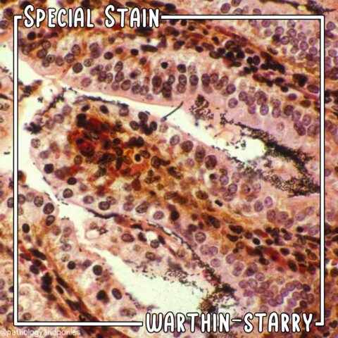
Today’s special stain is 𝐌𝐚𝐬𝐬𝐨𝐧’𝐬 𝐭𝐫𝐢𝐜𝐡𝐫𝐨𝐦𝐞!
𝐖𝐡𝐚𝐭 𝐢𝐬 𝐚 𝐌𝐚𝐬𝐬𝐨𝐧’𝐬 𝐭𝐫𝐢𝐜𝐡𝐫𝐨𝐦𝐞 𝐬𝐭𝐚𝐢𝐧?
Trichrome stains are primarily used to identify 𝐜𝐨𝐥𝐥𝐚𝐠𝐞𝐧 (a structural protein), bone or 𝐟𝐢𝐛𝐫𝐢𝐧 (the protein that forms blood clots). In general, collagen and bone are highlighted blue-green, while fibrin is highlighted in red.
This image is a case of 𝐞𝐧𝐝𝐨𝐜𝐚𝐫𝐝𝐢𝐚𝐥 𝐟𝐢𝐛𝐫𝐨𝐬𝐢𝐬 in the heart of a cat. In this condition, the 𝐞𝐧𝐝𝐨𝐜𝐚𝐫𝐝𝐢𝐮𝐦 (inner lining of the heart) becomes severely thickened with scar tissue. Scar tissue is primarily made up of collagen, which is why this heart is lined with bright blue on a trichrome stain!
Photo © Noah’s Arkive contributor Rech licensed under CC BY-SA 4.0.
