


Today’s path rounds are on 𝐫𝐢𝐜𝐤𝐞𝐭𝐬!
𝐖𝐡𝐚𝐭 𝐢𝐬 𝐢𝐭?
𝐑𝐢𝐜𝐤𝐞𝐭𝐬 is a disease of the skeleton in growing animals, that results in abnormal formation of bone at the growth plates, as well as poor quality bone in general.
𝐖𝐡𝐨 𝐠𝐞𝐭𝐬 𝐢𝐭?
Any species can get this! As mentioned previously, this disease specifically affects growing animals prior to the closure of their growth plates.
𝐖𝐡𝐚𝐭 𝐜𝐚𝐮𝐬𝐞𝐬 𝐢𝐭?
Basically, anything that can interfere with the mineralization of bone can cause rickets, but the majority of cases are due to dietary deficiencies in vitamin D or phosphorous.
Vitamin D deficiency is a major cause of rickets, particularly in animals kept indoors, due to lack of sunlight. Vitamin D plays a major role in calcium regulation by helping with calcium absorption in the diet. So, if an animal’s diet does not contain adequate vitamin D, the calcium levels in the blood stream decline, which stimulates the body to release calcium from the bones. This release of calcium is ultimately the underlying issue in rickets, because the bone cannot mineralize properly as it develops.
Phosphorous is another important factor in the formation of bone, because it is needed to form 𝐡𝐲𝐝𝐫𝐨𝐱𝐲𝐚𝐩𝐚𝐭𝐢𝐭𝐞, the major mineral in bones. Again, without adequate phosphorous levels, the bone cannot mineralize properly, causing rickets. This typically occurs in animals grazing on pastures where soil levels of phosphorous are low, and the diet is not otherwise balanced.
𝐖𝐡𝐲 𝐢𝐬 𝐭𝐡𝐢𝐬 𝐚 𝐩𝐫𝐨𝐛𝐥𝐞𝐦?
Well, having soft bones is not very conducive to living a long, happy and healthy life! These animals are generally very stiff and lame, and may even be reluctant to stand.
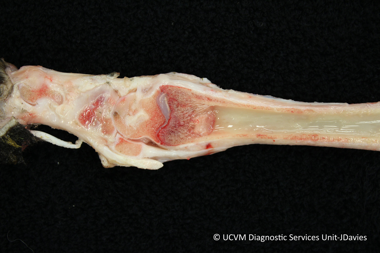
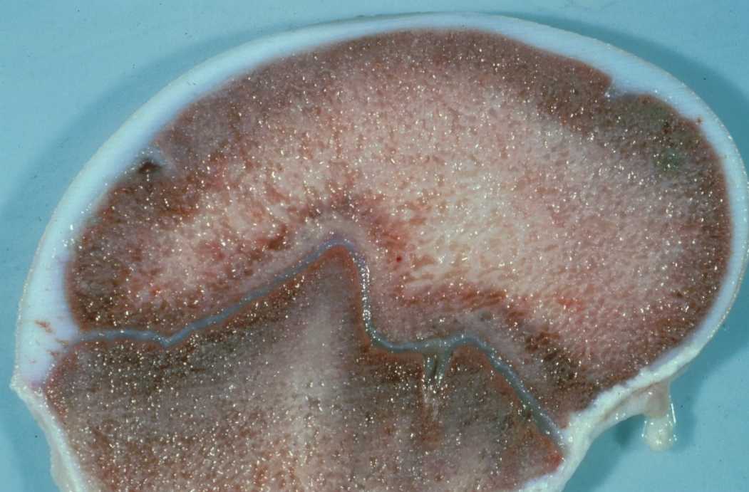
The bones often show irregular thickening of the growth plates, which can lead to deformities of the limbs as the growth plates become boney at different rates and thicknesses. This can further lameness and make animals extremely uncomfortable.
The bone itself can also have an irregular and disorganized appearance, reflecting its poor development. Because of how weak the bone structure is, these animals are significantly predisposed to bone fractures, even with mild traumas.
𝐇𝐨𝐰 𝐢𝐬 𝐢𝐭 𝐝𝐢𝐚𝐠𝐧𝐨𝐬𝐞𝐝?
Typically these lesions are initially diagnosed when the animal fractures a bone seemingly out of the blue. From there, the irregular growth pattern of the bone can be identified on X-ray, because they are less 𝐫𝐚𝐝𝐢𝐨𝐩𝐚𝐪𝐮𝐞 (white) than a normal bone.
𝐇𝐨𝐰 𝐢𝐬 𝐢𝐭 𝐭𝐫𝐞𝐚𝐭𝐞𝐝?
Typically, correcting the underlying dietary deficiency is the main treatment for rickets. If the animal is generally kept indoors, then letting it outside for frequently and for longer periods can also help. Usually animals have a good prognosis as long as the growth plates aren’t irreversibly damaged, and bone fractures can be prevented.
𝐏𝐡𝐨𝐭𝐨𝐬
1-3) Examples of bones displaying an irregular pattern in the 𝐭𝐫𝐚𝐛𝐞𝐜𝐮𝐥𝐚𝐞 (main structure) of the bone.
4-5) Two examples where the growth plate (the grey gelatinous looking thing) has also become irregular. Normally they are an even, thin line extending all the way across the bone!
6) A normal bone and growth plate you can use for comparison to the other bones! See how thin the growth plate is?
7) An interesting presentation of rickets often seen in humans and birds called a 𝐫𝐚𝐜𝐡𝐢𝐭𝐢𝐜 𝐫𝐨𝐬𝐚𝐫𝐲. What you’re seeing is enlargement of the 𝐜𝐨𝐬𝐭𝐨𝐜𝐡𝐨𝐧𝐝𝐫𝐚𝐥 𝐣𝐮𝐧𝐜𝐭𝐢𝐨𝐧 (the cartilage attachment between the ribs and the sternum) due to rickets. Crazy!
𝐒𝐨𝐮𝐫𝐜𝐞𝐬
Maxie, G. Jubb, Kennedy and Palmer’s Pathology of Domestic Animals, Volume 1. Sixth Edition.
Photos 2-3, 5-7 courtesy of Noah’s Arkive.
Photos 1 and 4 courtesy of University of Calgary Diagnostic Services Unit.


