Today’s path rounds are on 𝐩𝐲𝐨𝐭𝐡𝐨𝐫𝐚𝐱!
𝐖𝐡𝐚𝐭 𝐢𝐬 𝐢𝐭?
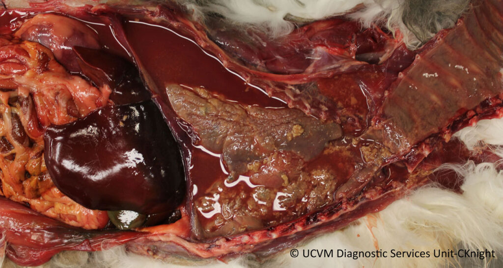
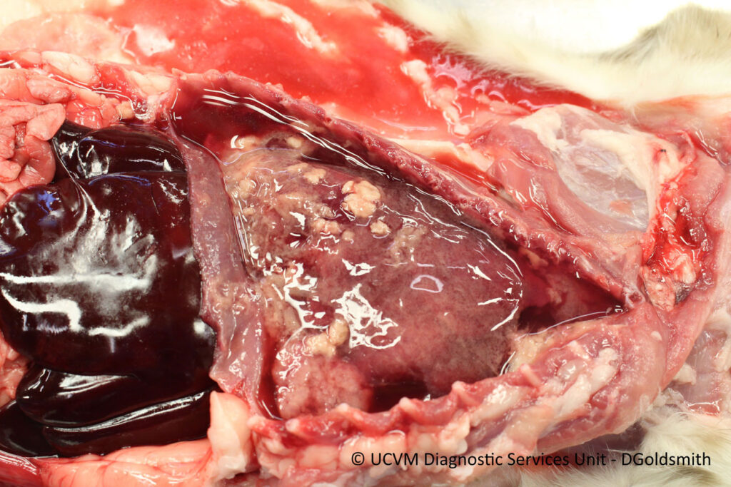
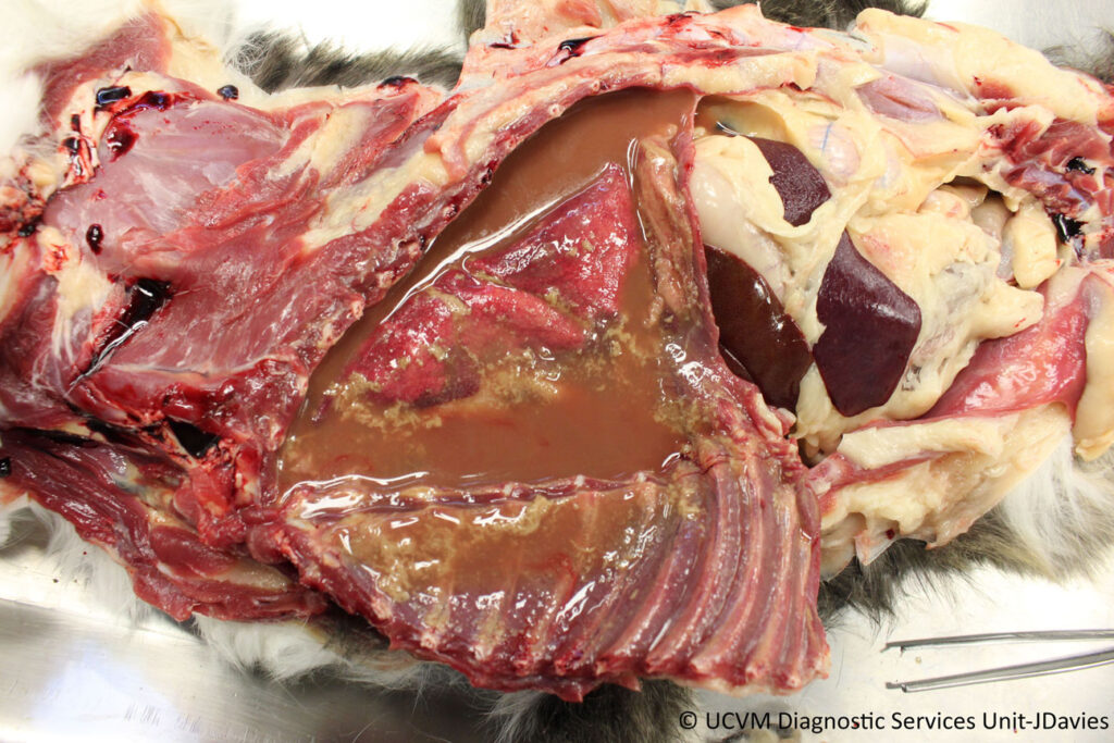
𝐏𝐲𝐨𝐭𝐡𝐨𝐫𝐚𝐱 is when pus (pyo-) accumulates within the thoracic cavity. Usually, this looks like large amounts of cloudy, yellow or red fluid that contains long strands of 𝐟𝐢𝐛𝐫𝐢𝐧 (one of the main clotting materials in the bloodstream).
𝐖𝐡𝐨 𝐠𝐞𝐭𝐬 𝐢𝐭?
Any species can get this! However, some of the most “classic” pyothorax lesions are seen in dogs and cats.
𝐖𝐡𝐚𝐭 𝐜𝐚𝐮𝐬𝐞𝐬 𝐢𝐭?
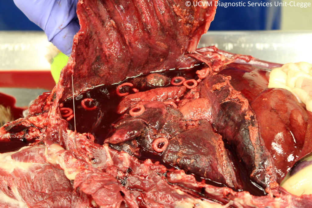
Anything that allows infectious agents, particularly bacteria, to reach the 𝐩𝐥𝐞𝐮𝐫𝐚, the lining of the lungs and thoracic cavity. This can be due to 𝐡𝐞𝐦𝐚𝐭𝐨𝐠𝐞𝐧𝐨𝐮𝐬 𝐬𝐩𝐫𝐞𝐚𝐝 (through the blood stream), penetrating wounds into the chest like bite wounds, abscesses in the lungs rupturing, or even foreign bodies in the esophagus or stomach perforating through the organ wall.
𝐖𝐡𝐲 𝐢𝐬 𝐭𝐡𝐢𝐬 𝐚 𝐩𝐫𝐨𝐛𝐥𝐞𝐦?
To make a long story short, the lungs do not like to be in water. They are not able to expand properly, leading to difficulty breathing. Because you have an infection running rampant as well, the animal will also have a fever, and may even develop 𝐬𝐞𝐩𝐭𝐢𝐜𝐞𝐦𝐢𝐚, where the bacteria enters the bloodstream and spreads around the body.
𝐇𝐨𝐰 𝐢𝐬 𝐢𝐭 𝐝𝐢𝐚𝐠𝐧𝐨𝐬𝐞𝐝?
Based on suspicion on clinical exam, the veterinarian will often take an X-ray or do an ultrasound. Both of these diagnostic methods will allow the veterinarian to see the fluid directly, furthering their suspicion. To confirm that it is a pyothorax instead of other types of fluid, they may do a 𝐭𝐡𝐨𝐫𝐚𝐜𝐨𝐜𝐞𝐧𝐭𝐞𝐬𝐢𝐬, where they put a needle into the thorax to extract some of the fluid. From there, they can assess the fluid to show the inflammatory component.
At necropsy, we can see the characteristic cloudy fluid to be able to make our initial diagnosis. Sometimes, the fluid is somewhat creamy looking and red tinged, and you can occasionally see clumps of yellow bacterial nests within the fluid. This leads to some pathologists calling it a “tomato soup with croutons” appearance. Gross.
𝐇𝐨𝐰 𝐢𝐬 𝐢𝐭 𝐭𝐫𝐞𝐚𝐭𝐞𝐝?
Drainage of fluid via thoracocentesis is the main treatment for this condition. Sometimes, this will need to be done multiple times. On top of this, the animal will need antibiotics to get the infection under control, and often an 𝐚𝐧𝐭𝐢𝐩𝐲𝐫𝐞𝐭𝐢𝐜 (a medication to reduce fever).
𝐏𝐡𝐨𝐭𝐨𝐬
1-3) Some examples of tomato soup with croutons pyothorax.
4) An example of pyothorax caused by an esophageal rupture after an obstruction. Those little weird donuts floating around are whatever the dog ate that caused the obstruction!
𝐒𝐨𝐮𝐫𝐜𝐞𝐬
Maxie, G. Jubb, Kennedy and Palmer’s Pathology of Domestic Animals, Volume 2. Sixth Edition.
Photos 1-4 courtesy of University of Calgary Diagnostic Services Unit.


