


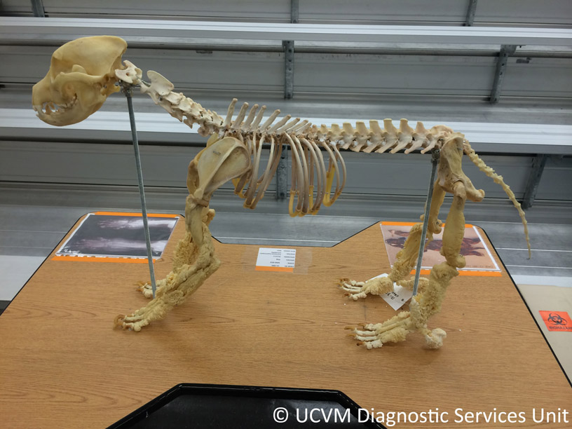
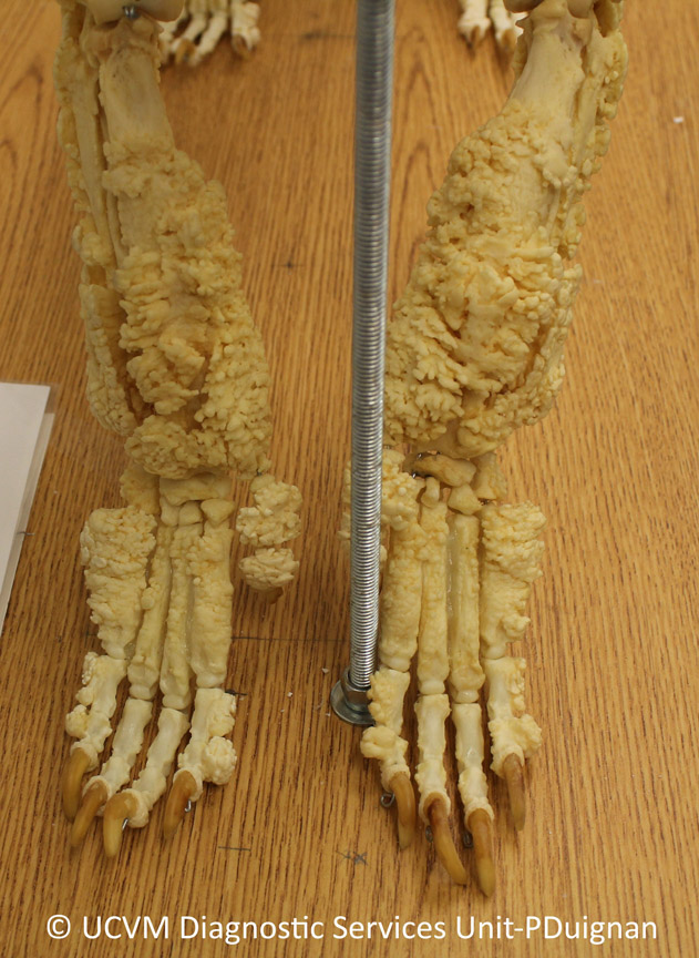
Today’s path rounds are on 𝐡𝐲𝐩𝐞𝐫𝐭𝐫𝐨𝐩𝐡𝐢𝐜 𝐨𝐬𝐭𝐞𝐨𝐩𝐚𝐭𝐡𝐲! This is one of my favourite diseases ![]() In fact, seeing the skeleton shown in these photos when I was at vet school was one of my big inspirations to go into pathology!
In fact, seeing the skeleton shown in these photos when I was at vet school was one of my big inspirations to go into pathology!
𝐖𝐡𝐚𝐭 𝐢𝐬 𝐢𝐭?
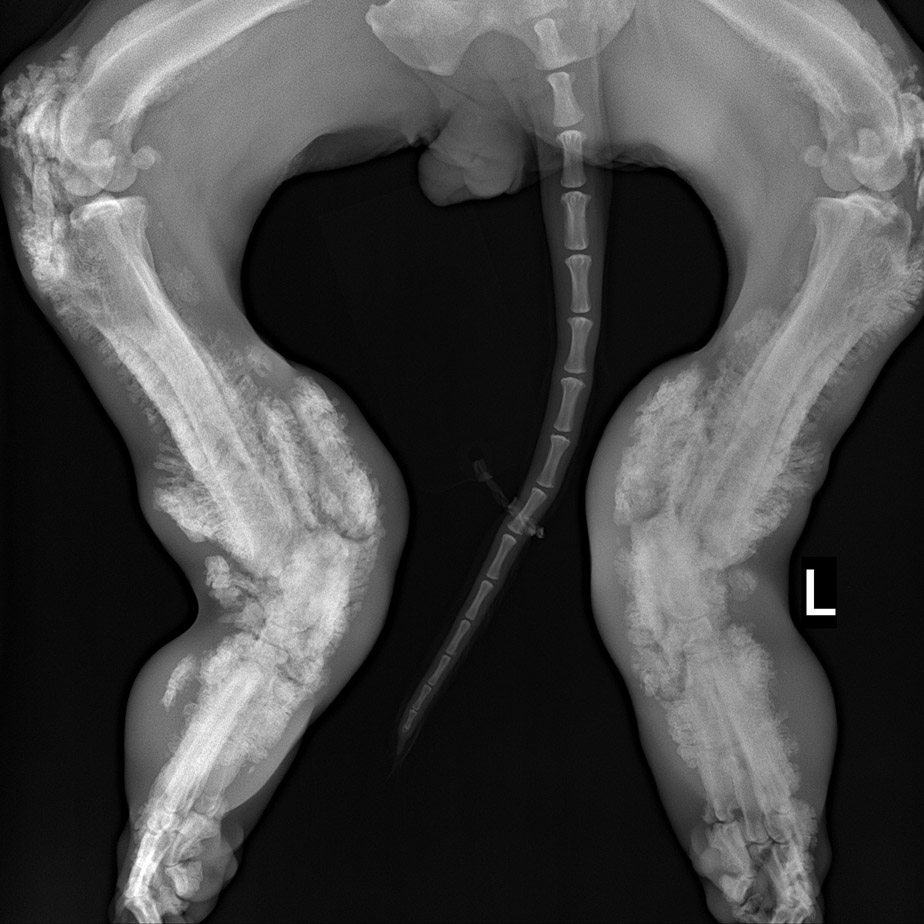
𝐇𝐲𝐩𝐞𝐫𝐭𝐫𝐨𝐩𝐡𝐢𝐜 𝐨𝐬𝐭𝐞𝐨𝐩𝐚𝐭𝐡𝐲 is characterized by 𝐩𝐞𝐫𝐢𝐨𝐬𝐭𝐞𝐚𝐥 𝐧𝐞𝐰 𝐛𝐨𝐧𝐞 𝐟𝐨𝐫𝐦𝐚𝐭𝐢𝐨𝐧, meaning proliferation of bone on the outside of existing bones. This proliferation affects the long bones of the limbs primarily.
𝐖𝐡𝐨 𝐠𝐞𝐭𝐬 𝐢𝐭?
This condition can be seen in many different species, but is most commonly reported in dogs.
𝐖𝐡𝐚𝐭 𝐜𝐚𝐮𝐬𝐞𝐬 𝐢𝐭?
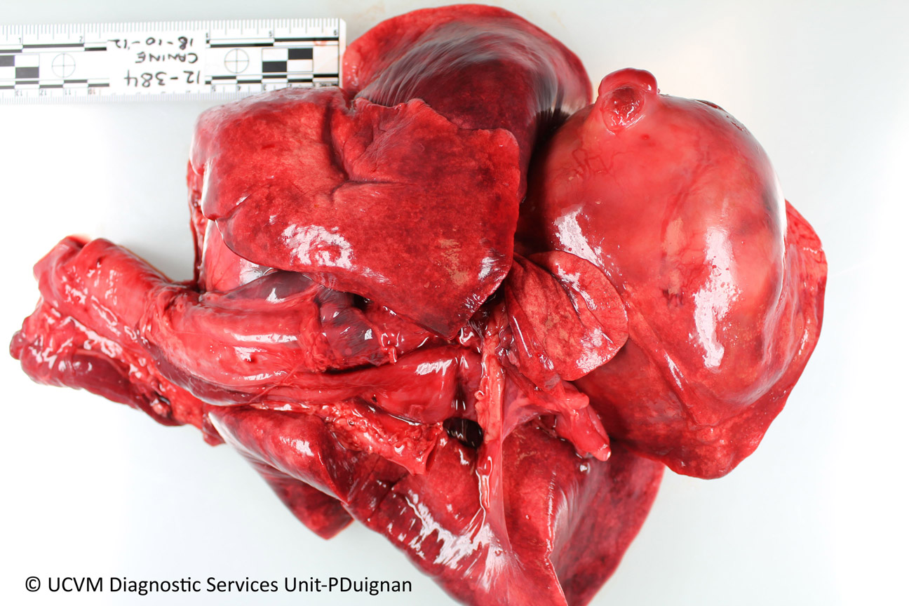
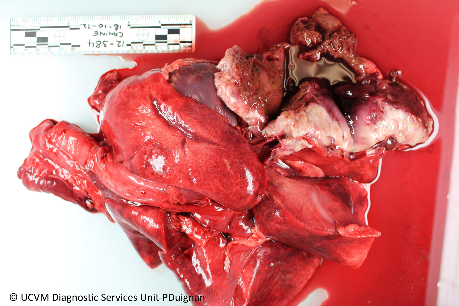
These proliferations are associated with chronic inflammatory or cancerous lesions in the thoracic cavity that are 𝐬𝐩𝐚𝐜𝐞-𝐨𝐜𝐜𝐮𝐩𝐲𝐢𝐧𝐠. How these space-occupying masses cause bone proliferation in the limbs is unknown! It has been suggested that thoracic lesions may release some type of hormone that that stimulates bone proliferation, or perhaps the lesion impedes on the vessels causing abnormal blood flow to the limbs.
𝐖𝐡𝐲 𝐢𝐬 𝐭𝐡𝐢𝐬 𝐚 𝐩𝐫𝐨𝐛𝐥𝐞𝐦?
These lesions are quite painful, but generally the bigger problem for the dog is whatever is going on in the thoracic cavity to cause the boney proliferation. In many cases, these space-occupying lesions are tumours, which is a sad time for the animal.
𝐇𝐨𝐰 𝐢𝐬 𝐢𝐭 𝐝𝐢𝐚𝐠𝐧𝐨𝐬𝐞𝐝?
The boney proliferation makes the legs look fat, which can be the first clue for the clinician that something is going on. From there, X-rays can show the boney proliferation, and X-rays or ultrasound can demonstrate the thoracic mass, confirming the diagnosis.
𝐇𝐨𝐰 𝐢𝐬 𝐢𝐭 𝐭𝐫𝐞𝐚𝐭𝐞𝐝?
Unfortunately there have not been many successful treatments identified. These animals are often euthanized due to the poor prognosis of their thoracic lesion.
𝐏𝐡𝐨𝐭𝐨𝐬
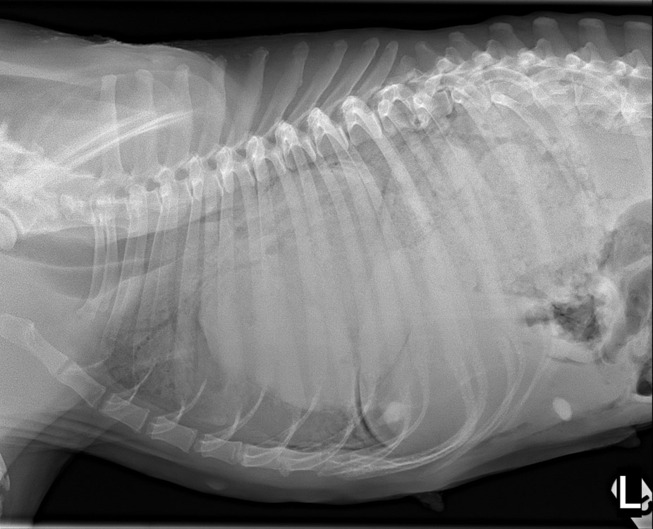
1-3) X-rays of a dog with hypertrophic osteopathy.
4) An X-ray of the same dog’s thorax showing a very opaque thorax! Normally the thorax is relatively clear.
5-6) The pulmonary mass that caused the hypertrophic osteopathy in this case.
7-8) The skeleton of the same dog after post-mortem. So crazy!!
𝐒𝐨𝐮𝐫𝐜𝐞𝐬
Maxie, G. Jubb, Kennedy and Palmer’s Pathology of Domestic Animals, Volume 1. Sixth Edition.
Photos 1-8 courtesy of University of Calgary Diagnostic Services Unit.


