
Today’s path rounds is on 𝐡𝐞𝐩𝐚𝐭𝐢𝐜 𝐥𝐢𝐩𝐢𝐝𝐨𝐬𝐢𝐬! Somewhat inspired by a case I had this week ![]()
𝐖𝐡𝐚𝐭 𝐢𝐬 𝐡𝐞𝐩𝐚𝐭𝐢𝐜 𝐥𝐢𝐩𝐢𝐝𝐨𝐬𝐢𝐬?
Hepatic lipidosis is basically when there is accumulation of 𝐭𝐫𝐢𝐠𝐥𝐲𝐜𝐞𝐫𝐢𝐝𝐞𝐬 (the smallest component of fat) within the liver cells.
𝐖𝐡𝐨 𝐠𝐞𝐭𝐬 𝐢𝐭?
Any species can get this, but cats in particular are very susceptible to it. The classic presentation of a hepatic lipidosis case is an obese female cat, that has some kind of stressor (e.g. moving, introduction of a new pet or family member, being accidentally locked somewhere without food etc) causing them to not eat.
𝐖𝐡𝐲 𝐢𝐬 𝐚𝐜𝐜𝐮𝐦𝐮𝐥𝐚𝐭𝐢𝐨𝐧 𝐨𝐟 𝐭𝐫𝐢𝐠𝐥𝐲𝐜𝐞𝐫𝐢𝐝𝐞𝐬 𝐚 𝐩𝐫𝐨𝐛𝐥𝐞𝐦?
On its own, this is not that big of a deal. In fact, one of the main functions of the liver is helping with triglyceride processing. However, very high amounts of triglycerides makes the cells more susceptible to disease, reduces their life-span and can impair their ability to repair themselves. So if another “secondary” problem comes along that also affects the liver, suddenly the liver has a BIG problem! Some examples of “secondary” problems are starvation, kidney disease, intestinal disease, tumours, diabetes, and more.
Without being treated, the mortality rate of hepatic lipidosis is very high. However with treatment, there is a good prognosis if it is caught early.
𝐖𝐡𝐚𝐭 𝐜𝐚𝐮𝐬𝐞𝐬 𝐢𝐭?
In cats, the accumulation of triglycerides likely has many contributing factors. Here are the two related factors we often think about:1) During periods of 𝐚𝐧𝐨𝐫𝐞𝐱𝐢𝐚 (animal is not eating), the body needs to retrieve fat out of storage in order to keep up with energy requirements. Remember how the liver helps with triglyceride processing? This means when all of that fat is pulled out of storage, the liver can get overwhelmed trying to keep up with all that processing! Thus, triglycerides start to accumulate within the liver cells, because they aren’t getting processed fast enough.2) Once the triglycerides are within the liver cells, the cell packages them into nice bundles called 𝐯𝐞𝐫𝐲 𝐥𝐨𝐰-𝐝𝐞𝐧𝐬𝐢𝐭𝐲 𝐥𝐢𝐩𝐨𝐩𝐫𝐨𝐭𝐞𝐢𝐧𝐬 (VLDLs). In order to make these nice packages, the liver cells need proteins, energy, and other components. If any of those “supplies” are low, which can also be caused by anorexia, then the cell can’t package things, causing triglycerides to accumulate.
𝐇𝐨𝐰 𝐢𝐬 𝐢𝐭 𝐝𝐢𝐚𝐠𝐧𝐨𝐬𝐞𝐝?
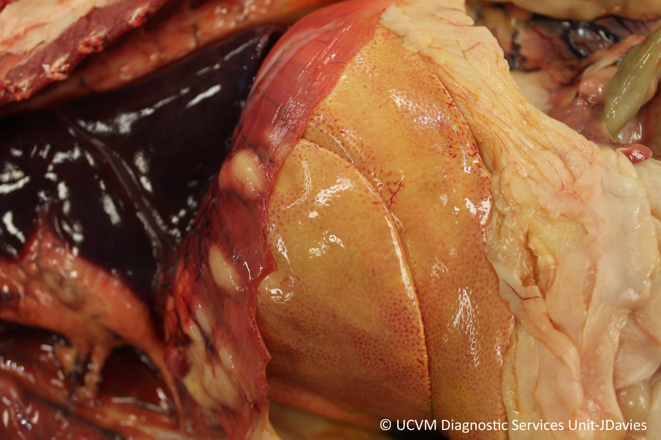
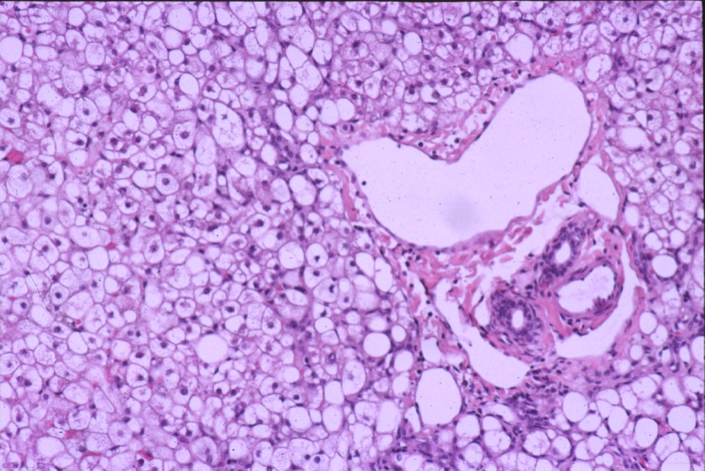
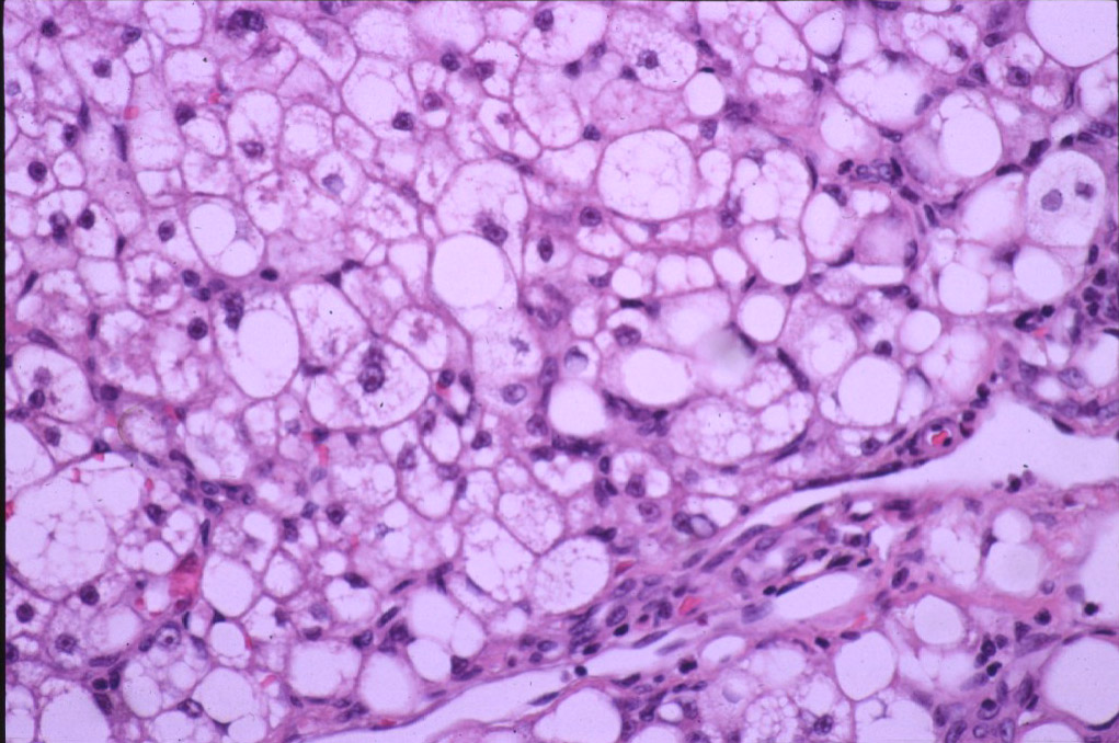
Cats presenting in a veterinary clinic will often have vomiting, anorexia, weakness and 𝐣𝐚𝐮𝐧𝐝𝐢𝐜𝐞 (yellowing of the tissues). Jaundice is a consequence of the liver cells not being able to excrete 𝐛𝐢𝐥𝐢𝐫𝐮𝐛𝐢𝐧, a yellow pigment that is formed when red blood cells are recycled or removed. Without the liver cells processing bilirubin to remove it from the blood, it accumulates in the tissues giving them a yellow tinge. On an ultrasound, the clinician may be able to see an enlarged liver, and on bloodwork, the liver values will be significantly elevated. At necropsy, the liver appears larger, yellow, and is more easily broken than a normal liver. They can also have more rounded edges than normal livers, and can feel kind of greasy. On histology, you can see large, clear 𝐯𝐚𝐜𝐮𝐨𝐥𝐞𝐬 (basically big storage bags within the cell) within the liver cells, which is where the triglycerides are stored.
𝐏𝐡𝐨𝐭𝐨𝐬
1) A very yellow liver in its normal position in the abdomen.
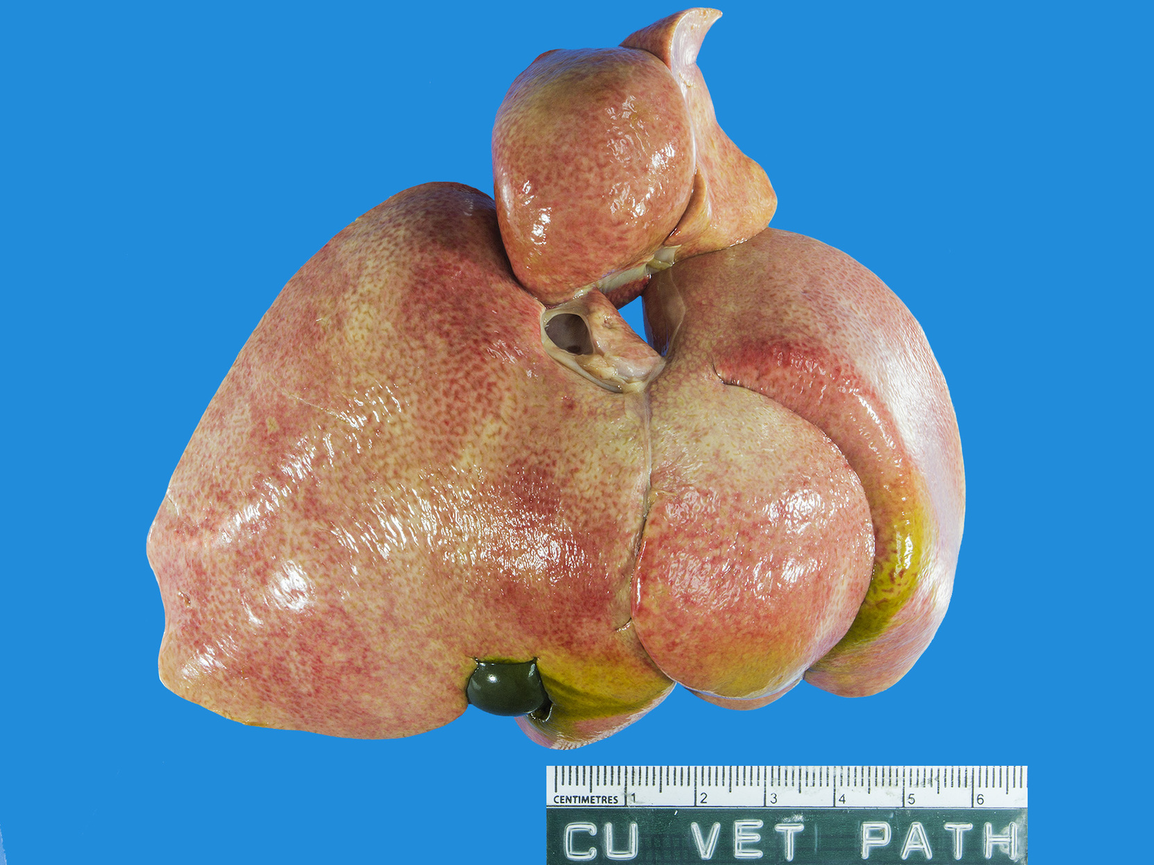
2) The 𝐜𝐫𝐚𝐧𝐢𝐚𝐥 (head-side) surface of a very yellow liver after being removed from the abdomen.
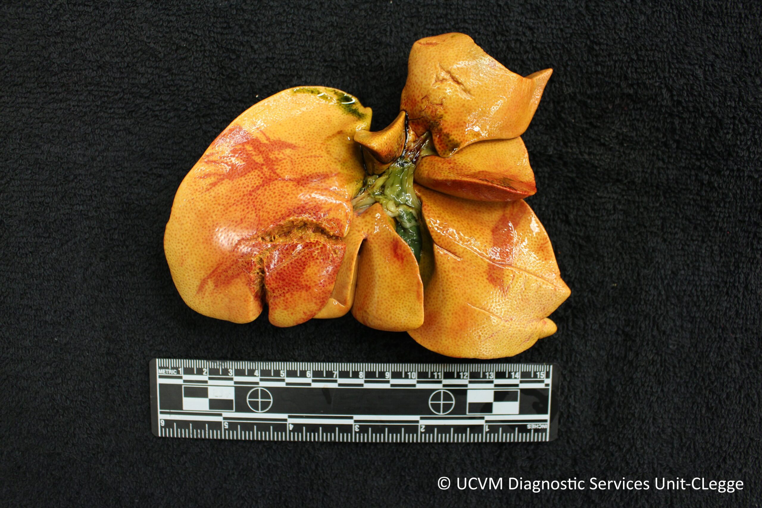
3) The 𝐜𝐚𝐮𝐝𝐚𝐥 (tail-side) surface of a very yellow liver after being removed from the abdomen. The green thing is the gall bladder!
4 and 5) The microscopic view of hepatic lipidosis. So many bubbles of triglyceride!
𝐒𝐨𝐮𝐫𝐜𝐞𝐬
Maxie, G. Jubb, Kennedy and Palmer’s Pathology of Domestic Animals, Volume 3. Sixth Edition.
Photos 1 and 3 courtesy of University of Calgary Veterinary Medicine Diagnostic Services Unit.
Photos 2, 4 and 5 courtesy of Noah’s Arkive.


