Today’s path rounds are on 𝐜𝐡𝐲𝐭𝐫𝐢𝐝𝐢𝐨𝐦𝐲𝐜𝐨𝐬𝐢𝐬!
𝐖𝐡𝐚𝐭 𝐢𝐬 𝐢𝐭?
𝐂𝐡𝐲𝐭𝐫𝐢𝐝𝐢𝐨𝐦𝐲𝐜𝐨𝐬𝐢𝐬 is a fungal infection of the skin of amphibians. Unfortunately, this disease has caused population declines in many wild amphibian species. Sometimes, it can even occur as outbreaks leading to a massive mortality event.
𝐖𝐡𝐨 𝐠𝐞𝐭𝐬 𝐢𝐭?
This disease affects any amphibian, but we most commonly think of it in frogs and salamanders.
𝐖𝐡𝐚𝐭 𝐜𝐚𝐮𝐬𝐞𝐬 𝐢𝐭?
The main cause of chytridiomyocosis are fungal species called 𝐁𝐚𝐭𝐫𝐚𝐜𝐡𝐨𝐜𝐡𝐲𝐭𝐢𝐮𝐦. This fungus infects the skin of the amphibians. Its life cycle begins with fungal bodies in the skin maturing to form 𝐳𝐨𝐨𝐬𝐩𝐨𝐫𝐚𝐧𝐠𝐢𝐚 containing many infected 𝐳𝐨𝐨𝐬𝐩𝐨𝐫𝐞𝐬 that have a flagella, allowing them to swim through water. These zoospores will infect other amphibians continuing the life cycle.
𝐖𝐡𝐲 𝐢𝐬 𝐭𝐡𝐢𝐬 𝐚 𝐩𝐫𝐨𝐛𝐥𝐞𝐦?
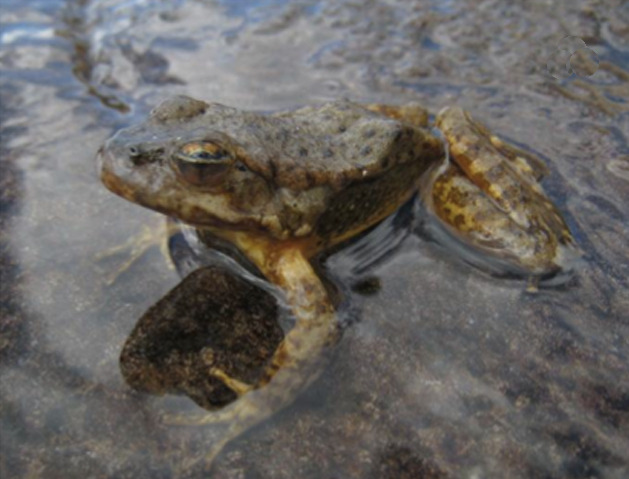
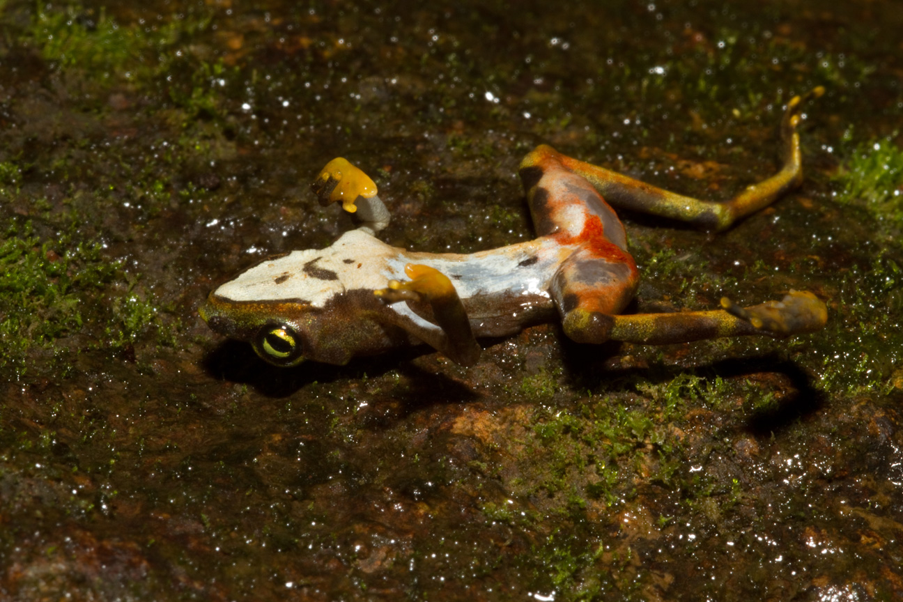
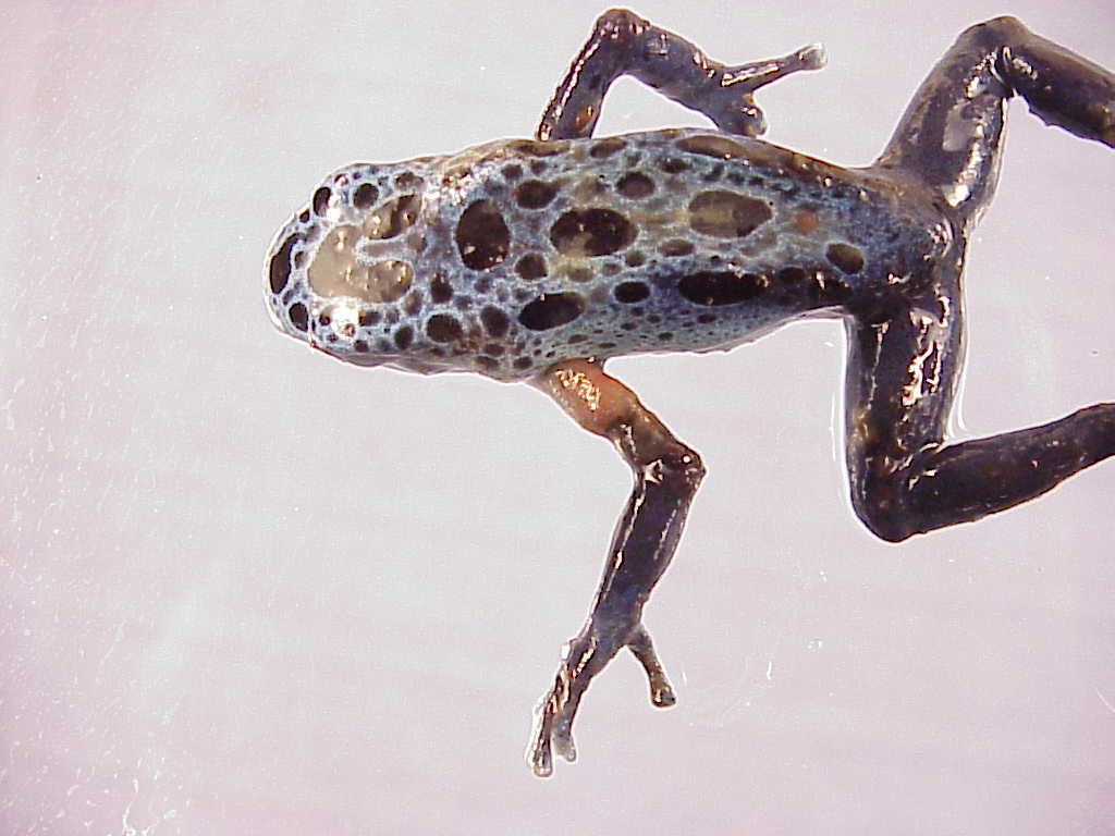
These fungi interrupt the normal function of the amphibian skin. This produces a rough skin texture, ulcers and a brownish or reddish skin discolouration. The skin of these amphibians also sheds incorrectly, called 𝐝𝐲𝐬𝐞𝐜𝐝𝐲𝐬𝐢𝐬. With this condition, the shedding skin comes off in small flakes rather than the typical large sheets.
The major consequence of this poor skin function is an inability to regulate their electrolytes, like sodium. These animals become 𝐡𝐲𝐩𝐨𝐧𝐚𝐭𝐫𝐞𝐦𝐢𝐜 (low sodium), 𝐡𝐲𝐩𝐨𝐤𝐚𝐥𝐞𝐦𝐢𝐜 (low potassium) and 𝐡𝐲𝐩𝐨𝐜𝐡𝐥𝐨𝐫𝐢𝐝𝐞𝐦𝐢𝐜 (low chloride), and ultimately die from cardiac arrest.
𝐇𝐨𝐰 𝐢𝐬 𝐢𝐭 𝐝𝐢𝐚𝐠𝐧𝐨𝐬𝐞𝐝?
The best way to diagnosis this disease is a skin biopsy, in order to visualize the fungi directly. Within the skin, a pathologist would be able to see 𝐡𝐲𝐩𝐞𝐫𝐤𝐞𝐫𝐚𝐭𝐨𝐬𝐢𝐬 (thickening of the skin layers) with many 𝐜𝐡𝐲𝐭𝐫𝐢𝐝 𝐭𝐡𝐚𝐥𝐥𝐢 (the skin-living form of the fungus). Often you can also see ulceration and 𝐧𝐞𝐜𝐫𝐨𝐬𝐢𝐬 (cell death) of the skin cells.
𝐇𝐨𝐰 𝐢𝐬 𝐢𝐭 𝐭𝐫𝐞𝐚𝐭𝐞𝐝?
The main treatment options being explored are temperature therapy and antifungal therapy. With temperature therapy, the amphibians are heated beyond the normal living temperature of the fungus. Antifungal therapy involves applying drugs toxic to the fungus to the amphibian, however this can be risky due to the highly absorptive nature of amphibian skin. Because of the high risks of treatment, these treatment protocols need to be completed under the supervision of a qualified veterinarian.
𝐏𝐡𝐨𝐭𝐨𝐬
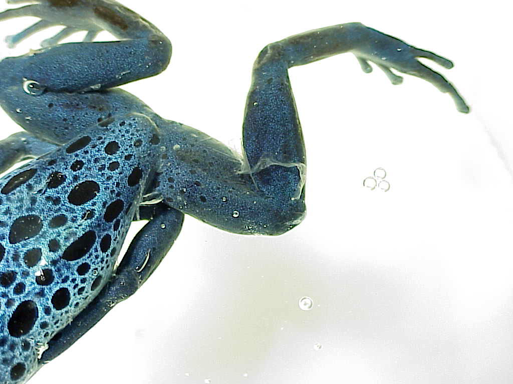
1-4) Sad frogs with chytridiomycosis, showing reddish brown skin and poor skin shedding.
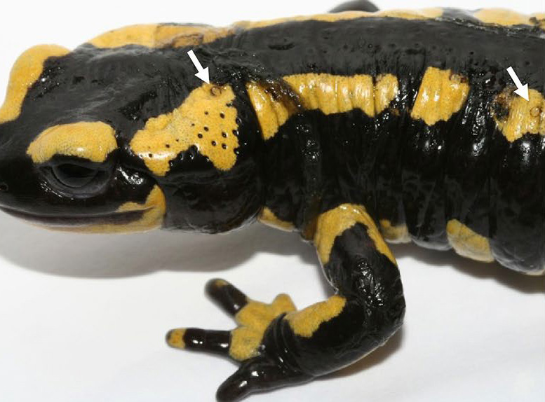
5) A salamander showing black to brown dots of chytridiomycosis.
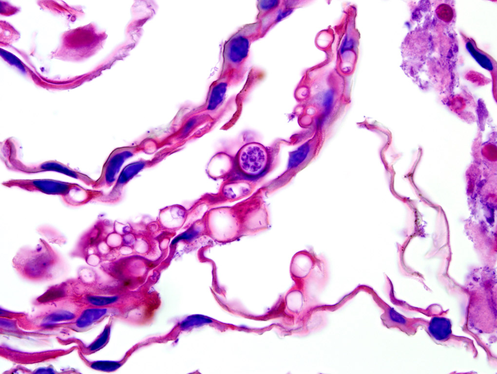
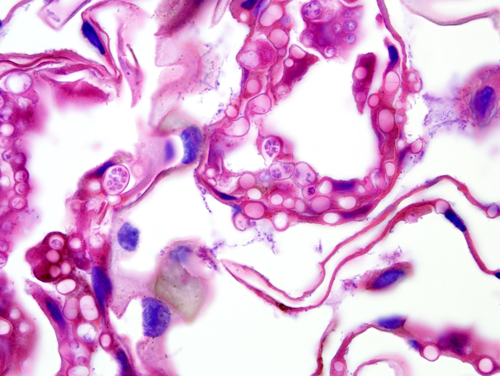
6-7) Absolutely beautiful microphotos of chytrid thalli in the skin of a frog!
𝐒𝐨𝐮𝐫𝐜𝐞𝐬
Terio, KA, McAloose, D, St. Leger, J. Pathology of Wildlife and Zoo Animals. 2018.
Photos 1-2, 6-7 © Noah’s Arkive contributors Terrell, Rech licensed under CC BY-SA 4.0.
Photo 3 © Wikimedia Commons contributor Gratwicke licensed under CC BY 2.0.
Photo 4 © Wikimedia Commons contributor Voyles licensed under CC BY 2.5.
Photo 5 © Wikimedia Commons contributor van Rooij licensed under CC BY 4.0.


