
It’s our 100th post! To celebrate I thought we could do something a little different… learning about what makes tissues different colours!
𝐇𝐨𝐰 𝐚𝐫𝐞 𝐜𝐨𝐥𝐨𝐮𝐫𝐬 𝐦𝐚𝐝𝐞?
Essentially, the colour of a tissue is produced by some commonly found cell or cell component that has a pigmentation. There are many different pigmented compounds throughout the body, and in some cases, they can even be deposited or accumulate excessively in disease conditions. This is just one of the features that a pathologist looks at 𝐠𝐫𝐨𝐬𝐬𝐥𝐲 (during necropsy) to help them understand what is happening in a tissue.
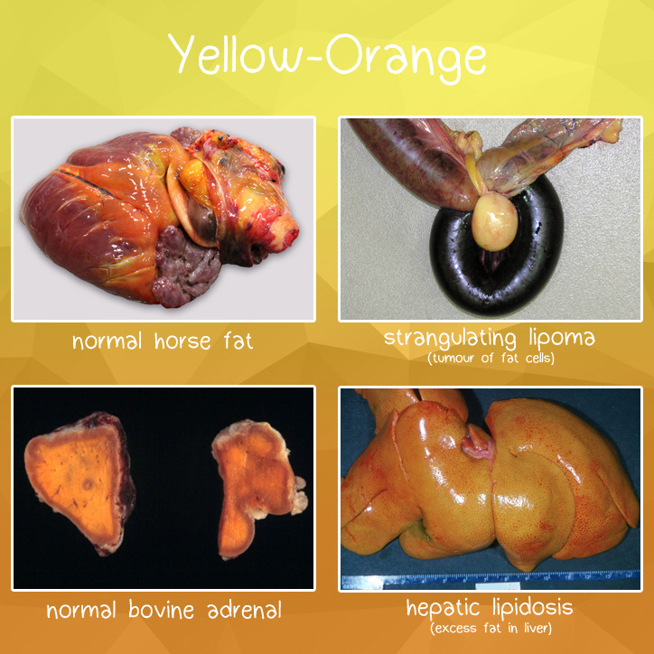
𝐘𝐞𝐥𝐥𝐨𝐰-𝐎𝐫𝐚𝐧𝐠𝐞
Yellow and orange colouring is most often caused by high levels of 𝐜𝐚𝐫𝐨𝐭𝐞𝐧𝐞𝐬, which is an important precursor to Vitamin A. The most common example of carotene pigmentation is… wait for it… 𝐜𝐚𝐫𝐫𝐨𝐭𝐬! Shocking. Carotenes are 𝐥𝐢𝐩𝐨𝐩𝐡𝐢𝐥𝐢𝐜, which means they primarily associate with fatty tissues in the body. This is what gives fat its yellow-ish tinge, and colours diseases with significant fat deposition yellow-orange as well.
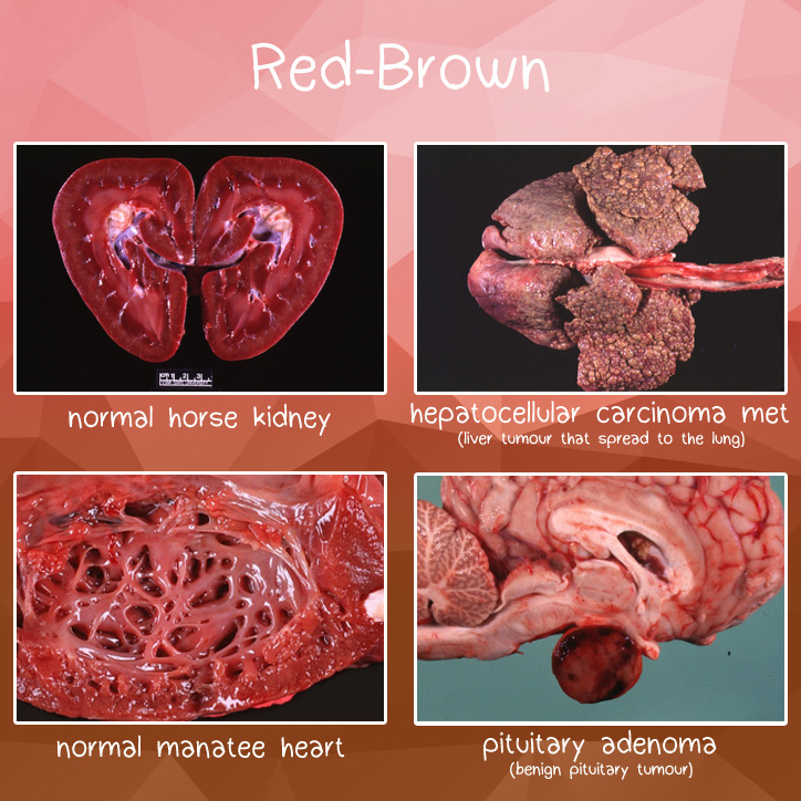
𝐑𝐞𝐝-𝐁𝐫𝐨𝐰𝐧
Red and brown are typically from 𝐡𝐞𝐦𝐞, which contains an iron molecule. In fact, heme is what makes blood red, as it is a component of 𝐡𝐞𝐦𝐨𝐠𝐥𝐨𝐛𝐢𝐧, the compound that gives red blood cells their oxygen carrying ability. Because blood flows through all tissues, this is what gives live tissue its pink to red colour. Some organs, like the liver and kidney, contain 𝐜𝐲𝐭𝐨𝐜𝐡𝐫𝐨𝐦𝐞𝐬, which are enzymes that help with metabolic functions, and also contain iron. These organs often are richly dark red to brown because of this additional pigment!
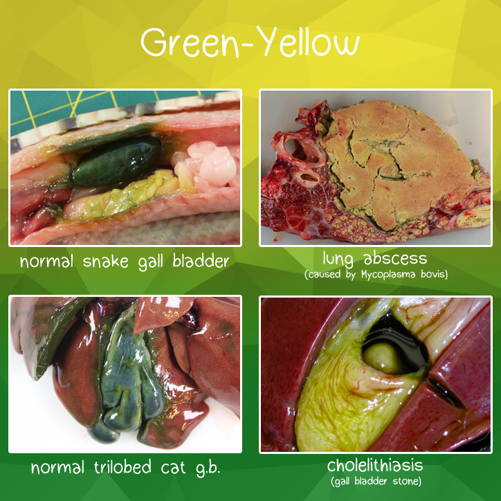
𝐆𝐫𝐞𝐞𝐧-𝐘𝐞𝐥𝐥𝐨𝐰
Green to yellow colouration is typically caused by 𝐛𝐢𝐥𝐢𝐫𝐮𝐛𝐢𝐧 and 𝐛𝐢𝐥𝐢𝐯𝐞𝐫𝐝𝐢𝐧, which are breakdown products of red blood cells. If you’ve ever wondered why a purple-red bruise turns yellow after a while, you can blame these two pigments for that! The best example of green in the body is the 𝐠𝐚𝐥𝐥𝐛𝐥𝐚𝐝𝐝𝐞𝐫, which is where all of this bilirubin is stored prior to excretion. Green to yellow can also be seen in abscesses, which is caused by infiltration of 𝐧𝐞𝐮𝐭𝐫𝐨𝐩𝐡𝐢𝐥𝐬 (major immune cell) that contains a yellow-green heme group.
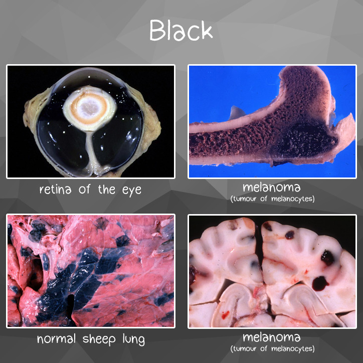
𝐁𝐥𝐚𝐜𝐤
Black colouration is produced by 𝐦𝐞𝐥𝐚𝐧𝐢𝐧, which is a pigment produced by 𝐦𝐞𝐥𝐚𝐧𝐨𝐜𝐲𝐭𝐞𝐬 mainly found in the skin. This is what gives skin its variety of shades! These pigments are also found in the retina of the eye, allowing a perfect environment for perception of light. Sometimes, the melanocytes can develop into tumours, producing richly black 𝐦𝐞𝐥𝐚𝐧𝐨𝐦𝐚𝐬 that can occur throughout the animal.
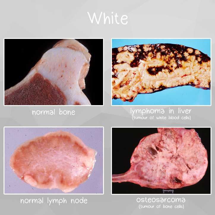
𝐖𝐡𝐢𝐭𝐞
There are actually a lot of different things that can make a tissue white, but the two main ones are 𝐜𝐚𝐥𝐜𝐢𝐮𝐦 and 𝐰𝐡𝐢𝐭𝐞 𝐛𝐥𝐨𝐨𝐝 𝐜𝐞𝐥𝐥𝐬. Calcium is most commonly found in bones and teeth, which is why these tissues are so strikingly white! However, calcium can also be deposited in tumours that produce bone or mineral as well. White blood cells themselves are actually white, which makes the 𝐥𝐲𝐦𝐩𝐡𝐨𝐢𝐝 𝐭𝐢𝐬𝐬𝐮𝐞 like our lymph nodes white as well. These densely packed white blood cells are also what make 𝐥𝐲𝐦𝐩𝐡𝐨𝐦𝐚𝐬 (tumours of white blood cells) white on cut section!Hope you guys enjoyed this brief tour of the different colours we see in pathology!
𝐒𝐨𝐮𝐫𝐜𝐞𝐬
Pina-Oviedo S, Ortiz-Hidalgo C, Ayala AG. Human Colors – the rainbow garden of pathology.
Photos © Noah’s Arkive contributors Gould, Larsen, Read, Render, Dick, Cho, Gross, Harrington, Nyo, Garlick, Hall, Vet Biosciences, Moore, Rech, Wright licensed under CC BY-SA 4.0.
Photos © University of Calgary Diagnostic Services Unit.


