Today’s path rounds are on 𝐬𝐭𝐫𝐚𝐧𝐠𝐮𝐥𝐚𝐭𝐢𝐧𝐠 𝐥𝐢𝐩𝐨𝐦𝐚𝐬! Inspired by a case I had last week ![]()
𝐖𝐡𝐚𝐭 𝐢𝐬 𝐢𝐭?
𝐒𝐭𝐫𝐚𝐧𝐠𝐮𝐥𝐚𝐭𝐢𝐧𝐠 𝐥𝐢𝐩𝐨𝐦𝐚𝐬 are one of the many causes of colic in horses. In this condition, there is a 𝐥𝐢𝐩𝐨𝐦𝐚 (a benign tumour of fat) that is 𝐩𝐞𝐝𝐮𝐧𝐜𝐮𝐥𝐚𝐭𝐞𝐝 (a long stalk with a mass on the end). Thanks to this long stalk, the mass can ends up wrapped around a piece of intestine, causing 𝐢𝐧𝐟𝐚𝐫𝐜𝐭𝐢𝐨𝐧 (complete loss of blood supply).
𝐖𝐡𝐨 𝐠𝐞𝐭𝐬 𝐢𝐭?
This condition is pretty much exclusive to horses. Typically it is teenaged or older horses that get this condition, because the mass takes time to develop.
𝐖𝐡𝐲 𝐢𝐬 𝐭𝐡𝐢𝐬 𝐚 𝐩𝐫𝐨𝐛𝐥𝐞𝐦?
Not having a blood supply to a tissue can cause a lot of issues. In this case, the blocked piece of intestine becomes 𝐞𝐝𝐞𝐦𝐚𝐭𝐨𝐮𝐬 (accumulation of excess fluid in the tissue), 𝐜𝐨𝐧𝐠𝐞𝐬𝐭𝐞𝐝 (non-flowing blood in the vessels) and 𝐡𝐞𝐦𝐨𝐫𝐫𝐡𝐚𝐠𝐢𝐜 (leakage of blood into the tissue). With time, the tissue of the intestine becomes 𝐧𝐞𝐜𝐫𝐨𝐭𝐢𝐜 (cell death), and may even rupture causing feed material to be released into the abdomen.
𝐖𝐡𝐲 𝐰𝐨𝐮𝐥𝐝 𝐭𝐡𝐞𝐬𝐞 𝐨𝐮𝐭𝐜𝐨𝐦𝐞𝐬 𝐡𝐚𝐩𝐩𝐞𝐧?
When the intestine becomes entrapped, the first structures to get compressed are the veins, because they have very thin and deformable walls. Veins normally drain blood out of an area, so if they become obstructed, no drainage can occur (congestion), despite the arteries still providing new blood to the tissue. This leads to increased pressure within the vessels as they try to cope with all the fluid. Eventually, the vessels become leaky, allowing fluid to escape, causing edema. If the vessels become significantly damaged, then red blood cells may be able to escape as well, causing hemorrhage.
𝐇𝐨𝐰 𝐢𝐬 𝐢𝐭 𝐝𝐢𝐚𝐠𝐧𝐨𝐬𝐞𝐝?
Typically horses with strangulating lipomas present with severe colic signs. As part of a diagnostic workup, the veterinarian may be able to feel dilated pieces of intestine on rectal examination, or visualize them using an ultrasound. These horses may also have 𝐫𝐞𝐟𝐥𝐮𝐱 (excessive fluid in the stomach) on 𝐧𝐚𝐬𝐨𝐠𝐚𝐬𝐭𝐫𝐢𝐜 𝐭𝐮𝐛𝐢𝐧𝐠 (passing a tube through the nose into the stomach), because feed material is unable to move through the intestines. If there is a rupture of the intestinal wall, the veterinarian may also see feed material on 𝐚𝐛𝐝𝐨𝐦𝐢𝐧𝐨𝐜𝐞𝐧𝐭𝐞𝐬𝐢𝐬 (sticking a needle into the abdomen to collect a fluid sample).
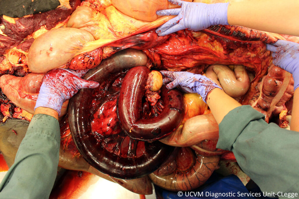
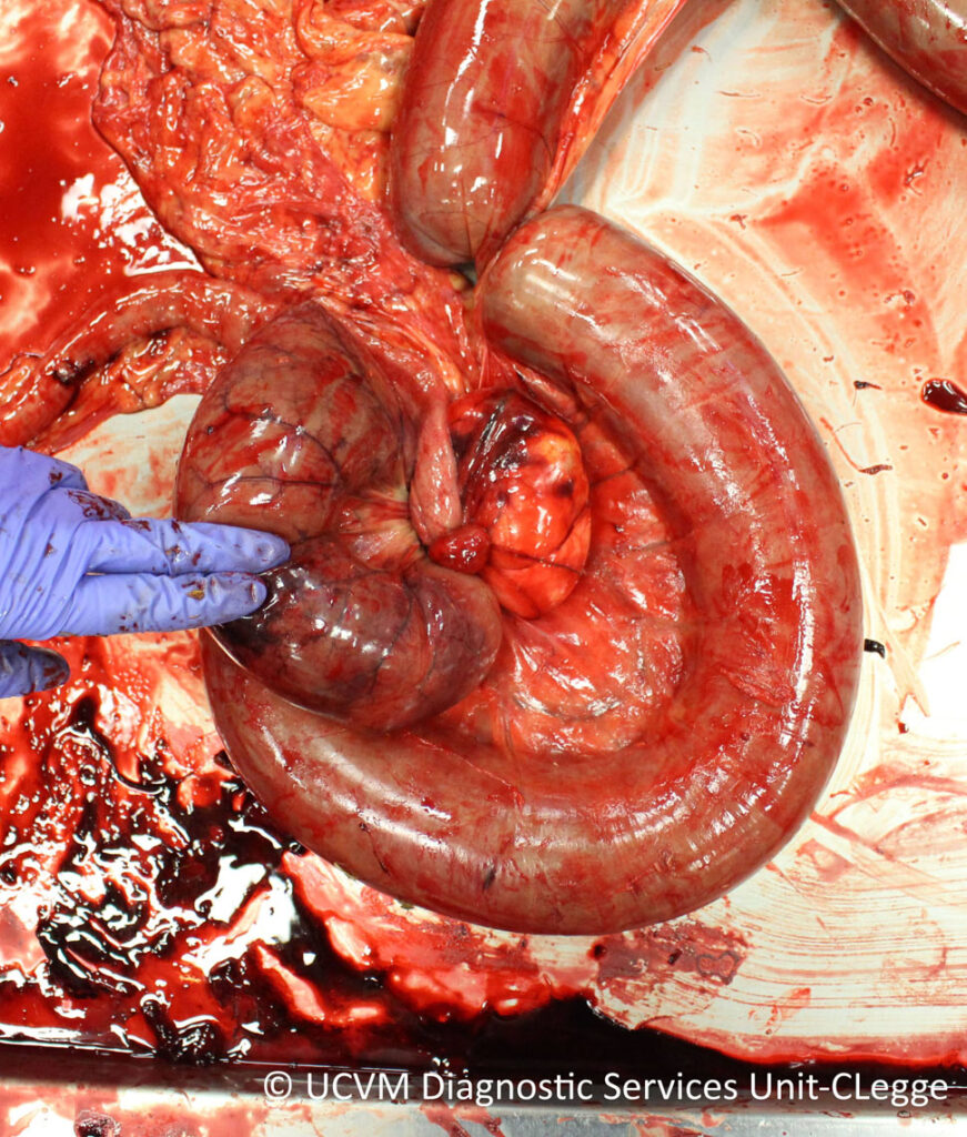
At necropsy, this condition is quite visually striking! Because the intestine that is entrapped becomes hemorrhagic, the pathologist can very quickly identify the affected piece of intestine due to its dark red to black coloration. From there, identifying the lipoma is easy, as you just have to follow the hemorrhagic intestine to its “source”.
𝐇𝐨𝐰 𝐢𝐬 𝐢𝐭 𝐭𝐫𝐞𝐚𝐭𝐞𝐝?
These have to be treated surgically, in order to remove the lipoma and put the intestines back into their correct positions. Often, horses will have to have a 𝐫𝐞𝐬𝐞𝐜𝐭𝐢𝐨𝐧 𝐚𝐧𝐝 𝐚𝐧𝐚𝐬𝐭𝐨𝐦𝐨𝐬𝐢𝐬 (removal of a piece of intestine) to remove the dead segment as well.
𝐏𝐡𝐨𝐭𝐨𝐬
1) The abdomen of a horse with a strangulating lipoma… can you see the affected piece of intestine?
2-4) Various examples of strangulating lipomas, with the lipomas and hemorrhagic intestine visible.
𝐒𝐨𝐮𝐫𝐜𝐞𝐬
Maxie, G. Jubb, Kennedy and Palmer’s Pathology of Domestic Animals, Volume 2. Sixth Edition.
Photos 1-3 courtesy of University of Calgary Diagnostic Services Unit.
Photo 4 courtesy of Noah’s Arkive.


