Today’s path rounds are on 𝐫𝐞𝐧𝐚𝐥 𝐢𝐧𝐟𝐚𝐫𝐜𝐭𝐬! No particular reason, just thought it would be a fun one.
𝐖𝐡𝐚𝐭 𝐢𝐬 𝐢𝐭?
𝐈𝐧𝐟𝐚𝐫𝐜𝐭𝐢𝐨𝐧 is when the blood supply to a tissue is blocked, causing that tissue to die. Infarcts in the kidney are somewhat unique because of their characteristic shape: each vessel in the kidney supplies a wedge shaped section of tissue, so when an infarct occurs, only that wedge of kidney is affected. So when you look at the kidney 𝐠𝐫𝐨𝐬𝐬𝐥𝐲 (without a microscope) you are able to see distinctive wedge shapes of infarcted tissue!
𝐖𝐡𝐨 𝐠𝐞𝐭𝐬 𝐢𝐭?
Any species can get this!
𝐖𝐡𝐚𝐭 𝐜𝐚𝐮𝐬𝐞𝐬 𝐢𝐭?
As mentioned previously, anything that causes a blockage of the blood supply to a tissue. Most commonly, these are 𝐞𝐦𝐛𝐨𝐥𝐢 (a chunk of something in the bloodstream, usually a tumor or bacterial colony) or 𝐭𝐡𝐫𝐨𝐦𝐛𝐢 (a blood clot). These get lodged in the small vessels supplying the kidney, causing a complete obstruction, and leading to the infarct. There are way too many causes of emboli/thrombi to list here!
𝐇𝐨𝐰 𝐝𝐨 𝐭𝐡𝐞𝐲 𝐜𝐡𝐚𝐧𝐠𝐞 𝐨𝐯𝐞𝐫 𝐭𝐢𝐦𝐞?
A neat thing about renal infarcts is you can tell how old they are, just by looking at them! When they first occur, they are often red due to 𝐜𝐨𝐧𝐠𝐞𝐬𝐭𝐢𝐨𝐧 (blood stagnant in the vessels because there is no outflow). The infarct will appear red for the first 2-3 days. Over the following 2-3 days, the blood sitting in this area will be broken down by 𝐧𝐞𝐮𝐭𝐫𝐨𝐩𝐡𝐢𝐥𝐬 (one of the immune system’s main clean-up cells), and the infarcted area will become white or pale. Over the next 1-2 weeks, the dead tissue is replaced by 𝐟𝐢𝐛𝐫𝐨𝐬𝐢𝐬(scar tissue), which is white and tends to be “depressed” and shrunken in appearance.
𝐖𝐡𝐲 𝐢𝐬 𝐭𝐡𝐢𝐬 𝐬𝐢𝐠𝐧𝐢𝐟𝐢𝐜𝐚𝐧𝐭?
If the tissue is infarcted and dead/dying, then it obviously can’t perform normal kidney tissue functions! Single, small infarcts aren’t usually an issue, because you can lose up to 70% of your renal tissue function before you start to show signs of renal failure. On necropsy, we would call these types of infarcts an 𝐢𝐧𝐜𝐢𝐝𝐞𝐧𝐭𝐚𝐥 𝐟𝐢𝐧𝐝𝐢𝐧𝐠, meaning that it was found but wasn’t a significant cause of disease. However, if there are several, large infarcts, the animal may enter renal failure, and then the infarcts would be a very significant finding!
𝐏𝐡𝐨𝐭𝐨𝐬
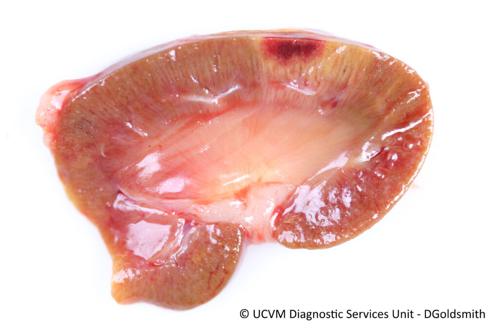
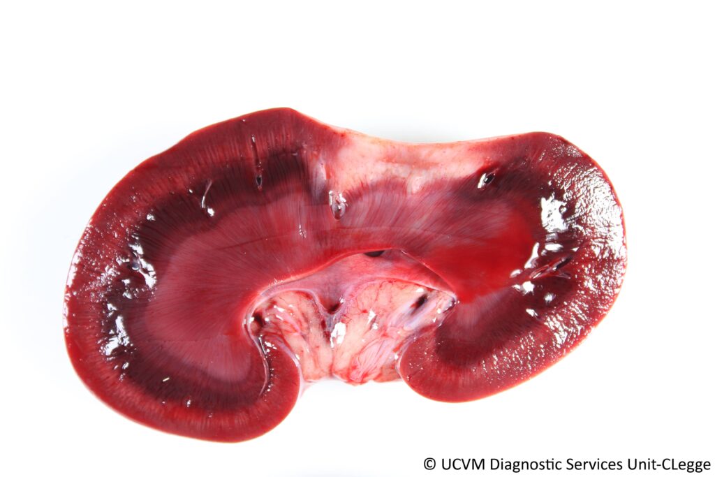
1) An 𝐚𝐜𝐮𝐭𝐞 (just happened!) renal infarct, showing the dark red coloration due to congestion.
2) A 𝐬𝐮𝐛𝐚𝐜𝐮𝐭𝐞 (happened a little while ago) renal infarct, showing it progressing to a pale tan color, still with a central area of red congestion.
3) A 𝐜𝐡𝐫𝐨𝐧𝐢𝐜 (happened a long time ago) renal infarct, showing a white, shrunken scar.
4) Renal infarcts can be seen even from the outside of the kidney! If this kidney looks weird to you, that’s because it kind of is. Cattle have lobulated kidneys, where each kidney is made up of a bunch of smaller “traditional” kidneys. Neat!
𝐒𝐨𝐮𝐫𝐜𝐞𝐬
Maxie, G. Jubb, Kennedy and Palmer’s Pathology of Domestic Animals, Volume 2. Sixth Edition.
Photos courtesy of University of Calgary Veterinary Medicine Diagnostic Services Unit.


