Heart failure is defined as when the heart’s pumping fails to meet the blood and oxygen demands of the tissues. Ultimately, this failure can be caused by decreased cardiac output, or decreased blood return. We most commonly think of the second mechanism when discussing heart failure, as this is the pathogenesis of congestive heart failure. However, it is important to note that low output heart failure can occur as well.
Failure of one side of the heart versus the other can also lead to different systemic consequences for the animal. So, it is important to distinguish between left sided and right sided heart failure. If both issues are occurring, we can call this bilateral heart failure.
To help distinguish between these types of heart failure, we will first focus on one side of the heart, then the other.
Table of Contents
Right-Sided Heart Failure
The job of the right side of the heart is to pull blood from the venous circulation, and pump it towards the lungs for oxygenation. Understanding this basic job is essential for understanding the effects of right-sided heart failure.
When the right side of the heart fails, the venous circulation is not cycled back into arterial circulation at a fast enough rate. This leads to accumulation of blood in the systemic veins, which is referred to as passive congestion. Clinically, you can see this congestion in the form of jugular vein distention, or even a jugular pulse. Internally, passive congestion affects the liver.
In the liver, we classically see “nutmeg liver”, or an enhanced lobular pattern, as the hallmark of chronic passive congestion. With right-sided heart failure, the centrilobular veins in the middle of the liver lobules aren’t drained properly, leading to congestion and dilation of the smaller vessels that would normally drain into it. Grossly, this makes the central portions of the lobule red. In the periportal regions, the cells experience hypoxia because fresh, oxygenated blood can’t make it into the congested lobule. This disrupts the cell’s normal fat processing, leading to fat accumulation. Grossly, this makes the periphery of the lobules look yellow. When these two processes are combined, it makes the liver look like a nutmeg!

© Jakowski licensed under CC BY-SA 4.0.
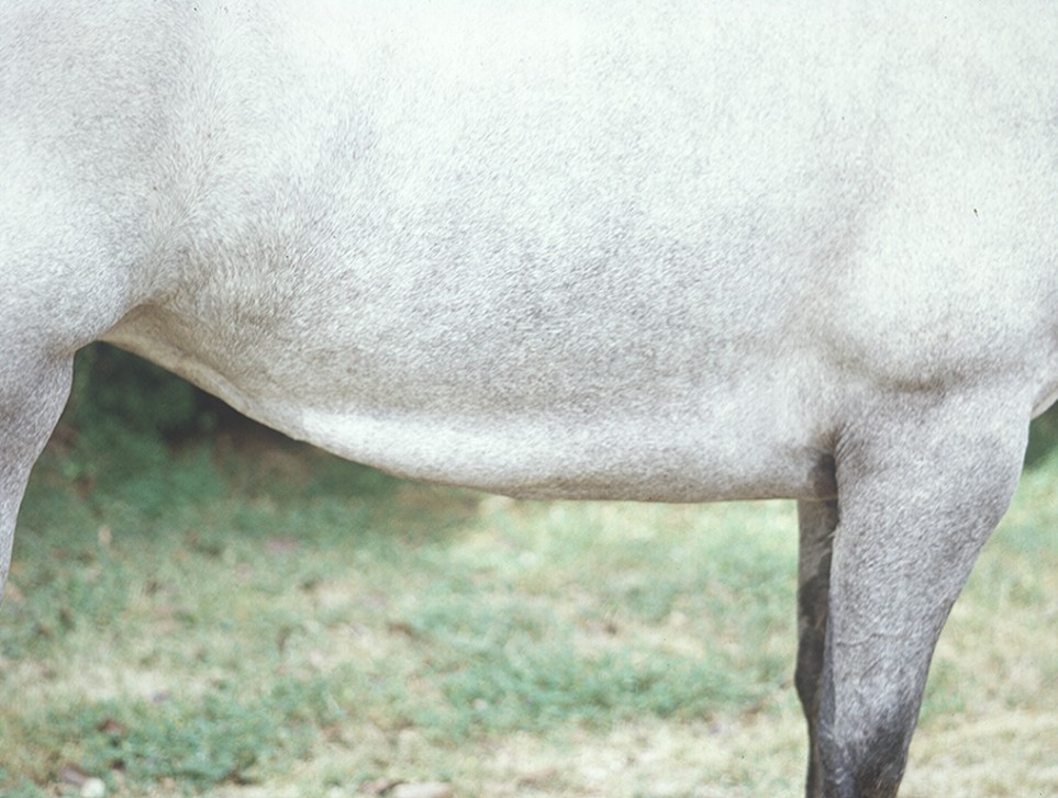
Although veins are very stretchy, they aren’t meant to hold all of the animal’s blood at once. Congestion increases the hydrostatic pressure within the vessels, which pushes more fluid out of the vessels than normal. Clinically, this change is probably the most obvious sign of right-sided heart failure! The increased pressure can lead to peripheral edema, ascites and pericardial effusion. Typically, dogs and cats are more likely to develop ascites, and horses and cows are more likely to develop peripheral edema.

© Harrington licensed under CC BY-SA 4.0.

© Crowell licensed under CC BY-SA 4.0.
Left-Sided Heart Failure
Similar to right-sided heart failure, the outcomes of left-sided heart failure are easy to identify if you know the job of the left side of the heart. This side of the heart pulls blood from the lungs and pumps it into arterial circulation. When the left side of the heart fails, there are two possible outcomes: congestion of the lungs, and failure to maintain cardiac output to the arterial circulation.
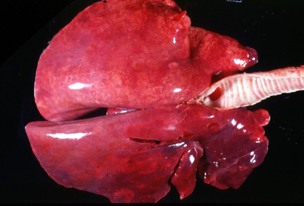
Congestion of the lungs occurs by a similar process to passive congestion of the liver. Put simply, the heart isn’t pulling enough blood from the lung circulation, causing it to accumulate in the pulmonary veins. This initially causes pulmonary congestion, which appears as very dark red lungs grossly.
Over time, the hydrostatic pressure in the veins will increase to the point where the vessels become leaky, leading to pulmonary edema. Grossly, these lungs appear enlarged and are very moist, and you may see clear fluid seep from the tissue when squeezed.
A brief note on cats:
Cats often present with pleural effusion with left-sided heart failure. The exact reason is unknown, but something to keep in mind!


© Crowell licensed under CC BY-SA 4.0.
Clinically, both of these conditions lead to tachypnea and dyspnea. Often, these animals will be exercise intolerant. Some dogs may have a cough associated with pulmonary edema.
The maintenance of arterial circulation by the heart is absolutely critical, particularly for the brain. Animals with left-sided heart failure can have their cardiac output compromised, meaning that there is a reduction in the amount of blood being pumped into circulation with each heartbeat. Because the brain relies on the blood’s oxygen stores, these animals may present with syncope, a transient loss of consciousness. They may also be generally depressed and lethargic due to inadequate delivery of glucose to the tissues. Clinically, you may also see hypotension in these animals.
Bilateral Heart Failure
With bilateral heart failure, the heart is just having a sad time in general. So, often clinical signs and gross lesions of both right and left-sided heart failure are present.
Comparison Table
| Type of Heart Failure | Congestion Location | Clinical Signs | Gross Lesions |
|---|---|---|---|
| Right-sided heart failure | Liver | Jugular vein distention Peripheral edema | Ascites “Nutmeg liver” Pericardial effusion |
| Left-sided heart failure | Lungs | Tachypnea and dyspnea Exercise intolerance Syncope Hypotension | Pulmonary congestion and edema Pleural effusion |
| Bilateral heart failure | Both liver and lungs | All of the above | All of the above |
Check Your Understanding
Zachary JF. Pathologic Basis of Veterinary Disease, Sixth Edition.
Kittleson MD. Heart Failure. Merck Veterinary Manual 2015.


