Today’s disease is 𝐦𝐞𝐥𝐚𝐧𝐨𝐦𝐚, also inspired by a recent case!
𝐖𝐡𝐚𝐭 𝐢𝐬 𝐢𝐭?
Melanomas are tumours that come from 𝐦𝐞𝐥𝐚𝐧𝐨𝐜𝐲𝐭𝐞𝐬, which are a type of cell primarily found in the skin. Melanocytes produce 𝐦𝐞𝐥𝐚𝐧𝐢𝐧, a dark pigment, that helps protect the skin cells from UV light.
𝐖𝐡𝐨 𝐠𝐞𝐭𝐬 𝐢𝐭?
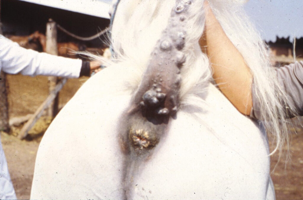
Pretty much any species can get it, however we most commonly see it in horses. In fact, it is one of the most common skin tumours seen in horses! Grey horses are predisposed to melanomas, with some studies showing up to 80% of older horses have at least one melanoma. They are most commonly found on the 𝐩𝐞𝐫𝐢𝐧𝐞𝐚𝐥 𝐫𝐞𝐠𝐢𝐨𝐧 (around the anus) and the underside of the tail, however they can be found anywhere on the body.
𝐇𝐨𝐰 𝐢𝐬 𝐢𝐭 𝐝𝐢𝐚𝐠𝐧𝐨𝐬𝐞𝐝?
A veterinarian can typically diagnose a melanoma based on the appearance of the tumour, and the location. If they are uncertain, they can take a 𝐛𝐢𝐨𝐩𝐬𝐲 (a sample of tissue) or an 𝐚𝐬𝐩𝐢𝐫𝐚𝐭𝐞 (a sample of cells) and send it to a pathologist for analysis.
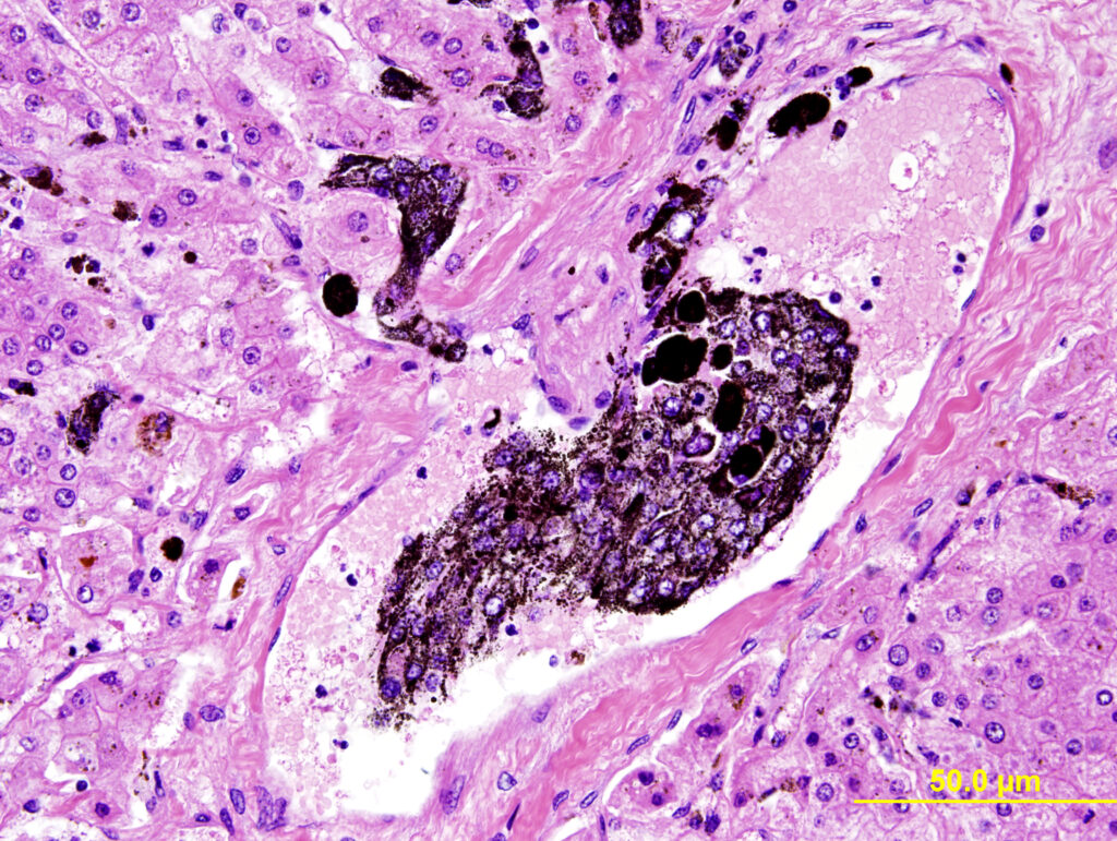
On biopsy or aspirate, melanoma cells are easily identified due to their characteristic dark pigment granules. These dark granules can sometimes cover up the nucleus of the cell though, which is where the pathologist will look to determine how aggressive the tumour is. To get around this, the pathologist might order a 𝐛𝐥𝐞𝐚𝐜𝐡𝐞𝐝 𝐬𝐥𝐢𝐝𝐞, which is exactly what it sounds like, because the bleach will remove the dark pigmentation of the granules. Then we can see the nucleus!
Some melanomas may be poorly pigmented, and without those easy-to-see dark granules it can be hard to tell what’s going on. So there are also special tests the pathologist can order to help confirm the diagnosis. One of those tests is 𝐌𝐞𝐥𝐚𝐧-𝐀, which is an 𝐢𝐦𝐦𝐮𝐧𝐨𝐡𝐢𝐬𝐭𝐨𝐜𝐡𝐞𝐦𝐢𝐬𝐭𝐫𝐲 𝐭𝐞𝐬𝐭 (𝐈𝐇𝐂). In this type of test, dyed antibodies against melanin pigment are applied to the section. When you look at the slide, anywhere that has melanin pigment shows up as coloured, allowing you to identify the pigment easily. Neat!
𝐇𝐨𝐰 𝐢𝐬 𝐢𝐭 𝐭𝐫𝐞𝐚𝐭𝐞𝐝?
These tumours are typically treated by surgical removal, and possibly chemotherapy. There has also been some work done to develop a “vaccine” for the tumour, primarily in dogs, which works by stimulating the immune system against the tumour cells.
𝐖𝐡𝐚𝐭 𝐢𝐬 𝐭𝐡𝐞 𝐩𝐫𝐨𝐠𝐧𝐨𝐬𝐢𝐬?
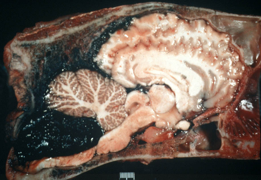
Unlike some tumour types, these tumours are typically slow growing and slow to metastasize. Therefore, they have a relatively good prognosis, but if left untreated, they may become more aggressive and spread quite quickly. If this occurs, horses may show severe signs of disease due to the tumours interfering with the normal function of organ systems. It is important to check your horses, especially grey horses, for lumps and bumps frequently!
𝐏𝐡𝐨𝐭𝐨𝐬
1) The classic appearance of melanomas on the underside of the tail of a grey horse.
2) An aspirate slide showing cells with characteristic dark pigment.
3) A biopsy slide showing the characteristic dark pigment. This slide happens to show the tumour cells within a blood vessel, on their way to metastasize elsewhere in the body.
4) Melanomas look black, even without a microscope! This photo shows what one of the nodules shown in the first photo might look like when cut.
5) Melanomas can metastasize to anywhere with a blood supply… even the brain!
𝐒𝐨𝐮𝐫𝐜𝐞𝐬
Phillips, J.C., Lembcke, L.M. Equine melanocytic tumours. Veterinary Clinics of North America: Equine Practice. 2013.
Photo 1-5 courtesy of Noah’s Arkive.


