Today’s path rounds are on 𝐥𝐲𝐦𝐩𝐡𝐚𝐧𝐠𝐢𝐞𝐜𝐭𝐚𝐬𝐢𝐚!
𝐖𝐡𝐚𝐭 𝐢𝐬 𝐢𝐭?
𝐋𝐲𝐦𝐩𝐡𝐚𝐧𝐠𝐢𝐞𝐜𝐭𝐚𝐬𝐢𝐚 is a big crazy word, but if you break it down it makes sense! Lymph- refers to the lymphatic system, angie- refers to vessels, and -ectasia refers to dilation. So putting it all together, lymphangiectasia is dilation of of the lymphatic vessels! Simple ![]() We typically see this disease in the intestine, but it can occur in other areas.
We typically see this disease in the intestine, but it can occur in other areas.
𝐖𝐡𝐨 𝐠𝐞𝐭𝐬 𝐢𝐭?
Any species can get this technically, however we most commonly see it in dogs. In particular, Yorkshire Terriers and Norwegian Lundehunds seem predisposed!
𝐖𝐡𝐚𝐭 𝐜𝐚𝐮𝐬𝐞𝐬 𝐢𝐭?
Basically lymphangiectasia is caused by anything obstructing the normal flow of lymph. So, we might consider things like inflammatory cells plugging up the vessel or even a tumour compressing the vessel. A large proportion of cases are considered 𝐢𝐝𝐢𝐨𝐩𝐚𝐭𝐡𝐢𝐜, meaning no obvious cause is identified, so those cases can be a bit of a mystery!
𝐖𝐡𝐲 𝐢𝐬 𝐭𝐡𝐢𝐬 𝐚 𝐩𝐫𝐨𝐛𝐥𝐞𝐦?
The lymphatics of the intestine are the major way that 𝐥𝐢𝐩𝐢𝐝 (fat) is absorbed by the body. When these lymphatics are blocked, the animal is unable to properly absorb this fat, and it remains in the intestine. The wall of the intestine is relatively leaky, and it wants to try and dilute out the intestinal contents to maintain balance. This causes a large amount of body fluid to enter the intestine to try and dilute the fat, leading to diarrhea and dehydration. The fluid lost into the intestine is often high in protein, leading to massive protein loss for the animal.
𝐇𝐨𝐰 𝐢𝐬 𝐢𝐭 𝐝𝐢𝐚𝐠𝐧𝐨𝐬𝐞𝐝?
These animals present with diarrhea, dehydration and weight loss, since they aren’t able to absorb fat properly. They may also have signs of vitamin deficiency, as some vitamins are 𝐟𝐚𝐭-𝐬𝐨𝐥𝐮𝐛𝐥𝐞, meaning they need to be dissolved in fat to be absorbed properly. On bloodwork, these animals will show a 𝐡𝐲𝐩𝐨𝐜𝐡𝐨𝐥𝐞𝐬𝐭𝐞𝐫𝐨𝐥𝐞𝐦𝐢𝐚 and 𝐡𝐲𝐩𝐨𝐩𝐫𝐨𝐭𝐞𝐢𝐧𝐞𝐦𝐢𝐚, essentially meaning low fat and protein in the blood. To confirm a diagnosis, a biopsy of the intestine can be submitted to a pathologist for examination.
𝐇𝐨𝐰 𝐢𝐬 𝐢𝐭 𝐭𝐫𝐞𝐚𝐭𝐞𝐝?
The first line of therapy, if possible, is removing the inciting cause. For example, removing the obstructing tumour, or resolving the inflammation. However, this is often not possible. Therefore, these animals are often managed on a low-fat, high-calorie diet. However, the overall prognosis for this condition is poor, so these animals are often euthanized before their quality of life declines further.
𝐏𝐡𝐨𝐭𝐨𝐬
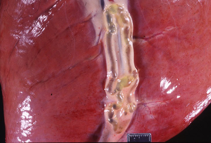
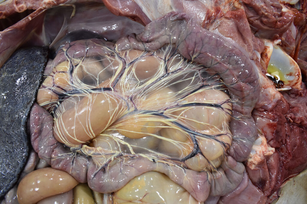
1-2) Examples of large lymphatic vessels showing dilation, in the heart and the 𝐦𝐞𝐬𝐞𝐧𝐭𝐞𝐫𝐲 (tissue supporting the intestines).
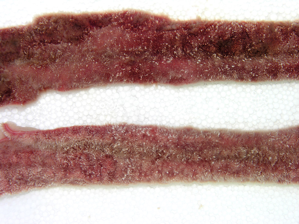
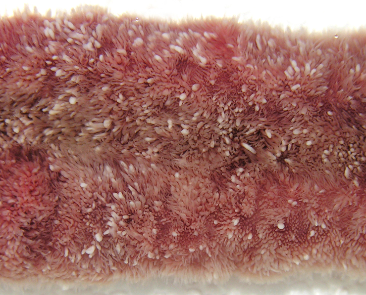
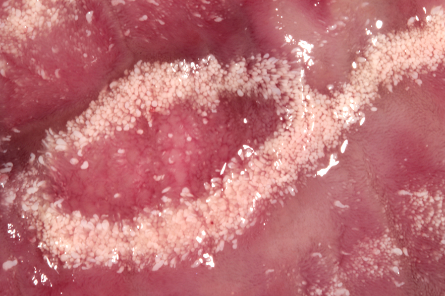
3-5) Close up photos of intestines with lymphangiectasia. The intestine is lined by little finger-like projections, each containing a large lymphatic vessel. In this disease, the dilation of the lymphatic vessel makes the little fingers look white, rather than their normal pink colour.
𝐒𝐨𝐮𝐫𝐜𝐞𝐬
Maxie, G. Jubb, Kennedy and Palmer’s Pathology of Domestic Animals, Volume 2. Sixth Edition.
Photos 1-5 © Noah’s Arkive contributor Rech, Read, Edwards, Neto licensed under CC BY-SA 4.0.

