Today’s path rounds are on 𝐢𝐧𝐭𝐮𝐬𝐬𝐮𝐬𝐜𝐞𝐩𝐭𝐢𝐨𝐧! This topic is related to a case I had recently.
𝐖𝐡𝐚𝐭 𝐢𝐬 𝐢𝐭?
𝐈𝐧𝐭𝐮𝐬𝐬𝐮𝐬𝐜𝐞𝐩𝐭𝐢𝐨𝐧 is when one piece of the intestine (called the 𝐢𝐧𝐭𝐮𝐬𝐬𝐮𝐬𝐜𝐞𝐩𝐭𝐮𝐦) telescopes into another piece of intestine (called the 𝐢𝐧𝐭𝐮𝐬𝐬𝐮𝐬𝐜𝐢𝐩𝐢𝐞𝐧𝐬), forming a multi-layered intestinal segment.
𝐖𝐡𝐨 𝐠𝐞𝐭𝐬 𝐢𝐭?
All species can get this!
𝐖𝐡𝐚𝐭 𝐜𝐚𝐮𝐬𝐞𝐬 𝐢𝐭?
This issue can be caused by linear foreign bodies like string or yarn in the intestine, parasites, previous intestinal surgery, inflammation of the intestine or even tumours. Basically, anything that can disrupt the normal gut motility of a section of intestine. When the gut stops moving in one area, the 𝐨𝐫𝐚𝐝 (closer to the mouth) segment of intestine continues to move, causing it to basically crawl into the non-motile intestine, forming the intussusception.
𝐖𝐡𝐲 𝐚𝐫𝐞 𝐭𝐡𝐞𝐲 𝐚 𝐩𝐫𝐨𝐛𝐥𝐞𝐦?
When a piece of intestine is pulled into another section of intestine, it compresses the veins that drain the internalized section. This causes 𝐜𝐨𝐧𝐠𝐞𝐬𝐭𝐢𝐨𝐧 (static, non-flowing blood in the vessels) and 𝐞𝐝𝐞𝐦𝐚 (excess fluid in the tissue) of the internalized section. Eventually, this leads to tissue death for the internalized section, potentially causing release of intestinal contents into the abdomen and causing a massive infection called 𝐩𝐞𝐫𝐢𝐭𝐨𝐧𝐢𝐭𝐢𝐬. Additionally, intestinal contents generally cannot move through an intussusception, causing severe abdominal pain for the animal.
𝐇𝐨𝐰 𝐢𝐬 𝐢𝐭 𝐝𝐢𝐚𝐠𝐧𝐨𝐬𝐞𝐝?
Intussusceptions can be diagnosed by ultrasound, where you can see a “target” like image made by the layering of the intestine. X-rays may also help, especially when the animal is given 𝐛𝐚𝐫𝐢𝐮𝐦 orally, as it shows up as white on an X-ray. With barium, you can see dilation of the intestine with backed up intestinal contents before the intussusception, and minimal intestinal contents after the intussusception.
𝐇𝐨𝐰 𝐢𝐬 𝐢𝐭 𝐭𝐫𝐞𝐚𝐭𝐞𝐝?
These are almost always treated surgically, by doing a 𝐫𝐞𝐬𝐞𝐜𝐭𝐢𝐨𝐧 𝐚𝐧𝐝 𝐚𝐧𝐚𝐬𝐭𝐨𝐦𝐨𝐬𝐢𝐬. This means that the affected piece of intestine is removed (resection) and then intestine is sutured back together without that affected part (anastomosis). Generally these surgeries are reasonably successful, especially if there is no peritonitis.
𝐏𝐡𝐨𝐭𝐨𝐬
1) A diagram showing the basic idea behind intussusception.
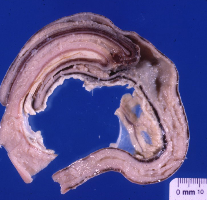
2) A cool 𝐬𝐚𝐠𝐢𝐭𝐭𝐚𝐥 (lengthwise) section of an intussusception showing how the intestine has moved into the rest of the intestine.
3) A cross-section of an intussusception showing the layers in a different way. Do you see why it looks like a target on ultrasound?
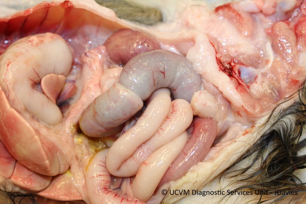
4-6) Photos of different intussusceptions, including one from a Ferruginous hawk (Photo 6)!
𝐒𝐨𝐮𝐫𝐜𝐞𝐬
Maxie, G. Jubb, Kennedy and Palmer’s Pathology of Domestic Animals, Volumes 2. Sixth Edition.
Photo 1 courtesy of Wikimedia Commons.
Photos 2-4 courtesy of Noah’s Arkive.
Photo 5-6 courtesy of University of Calgary Diagnostic Services Unit.
