


OLYMPUS DIGITAL CAMERA 
OLYMPUS DIGITAL CAMERA 
OLYMPUS DIGITAL CAMERA
Today’s path rounds are on 𝐜𝐡𝐨𝐫𝐝𝐨𝐦𝐚𝐬!
𝐖𝐡𝐚𝐭 𝐢𝐬 𝐢𝐭?
𝐂𝐡𝐨𝐫𝐝𝐨𝐦𝐚𝐬 are rare tumours that arise from remnants of the 𝐧𝐨𝐭𝐨𝐜𝐡𝐨𝐫𝐝, which are primitive cells leftover from embryonic development. These tumours tend to grow in the tail region in most species.
𝐖𝐡𝐨 𝐠𝐞𝐭𝐬 𝐢𝐭?
Technically any species can get this, however it is most commonly seen in ferrets!
𝐖𝐡𝐚𝐭 𝐜𝐚𝐮𝐬𝐞𝐬 𝐢𝐭?
The notochord is a group of primitive cells in the developing embryo that ultimately forms the spinal column. Some of these cells may not finish development, and end up hanging out in the areas in and around the spinal column. In rare cases, these cells may become 𝐧𝐞𝐨𝐩𝐥𝐚𝐬𝐭𝐢𝐜, undergoing uncontrolled replication and growth. This eventually forms the tumour that we identify as a chordoma!
𝐖𝐡𝐲 𝐢𝐬 𝐭𝐡𝐢𝐬 𝐚 𝐩𝐫𝐨𝐛𝐥𝐞𝐦?
As mentioned previously, chordomas typically arise on the tail, where they generally don’t cause too many issues. However, since they can technically arise anywhere along the spinal cord, some chordomas may cause significant issues. If they occur near the spinal cord, they can compress it and cause weakness and 𝐚𝐭𝐚𝐱𝐢𝐚 (irregular gait).
𝐇𝐨𝐰 𝐢𝐬 𝐢𝐭 𝐝𝐢𝐚𝐠𝐧𝐨𝐬𝐞𝐝?
Typically these are best diagnosed by looking at a 𝐛𝐢𝐨𝐩𝐬𝐲 (a sample of tissue) under a microscope. They are characteristically made up of 𝐩𝐡𝐲𝐬𝐚𝐥𝐢𝐟𝐞𝐫𝐨𝐮𝐬 𝐜𝐞𝐥𝐥𝐬, which are large cells that look like collections of bubbles! Sometimes they can be hard to differentiate from other types of tumours, so pathologists can stain the slide using antibodies against 𝐜𝐲𝐭𝐨𝐤𝐞𝐫𝐚𝐭𝐢𝐧 or 𝐯𝐢𝐦𝐞𝐧𝐭𝐢𝐧, which are both cell structural proteins.
𝐇𝐨𝐰 𝐢𝐬 𝐢𝐭 𝐭𝐫𝐞𝐚𝐭𝐞𝐝?
Because these tumours typically occur on the tail, they are usually easily removed by simply amputating that section of the tail. If they occur in other regions, they are difficult, if not impossible, to remove, so often these animals are euthanized if their quality of life is compromised.
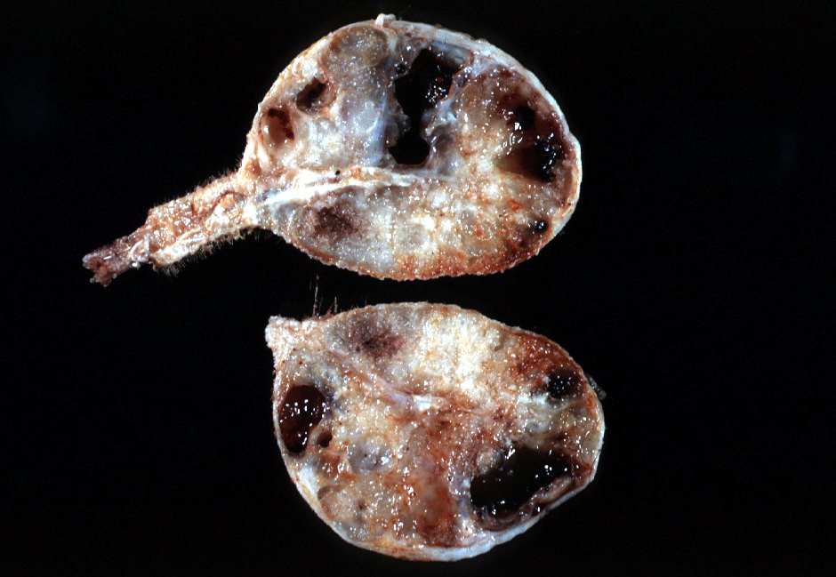
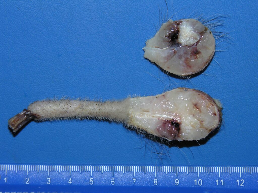
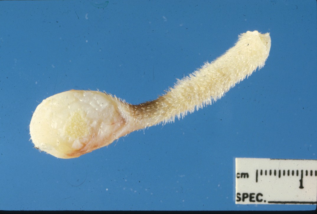
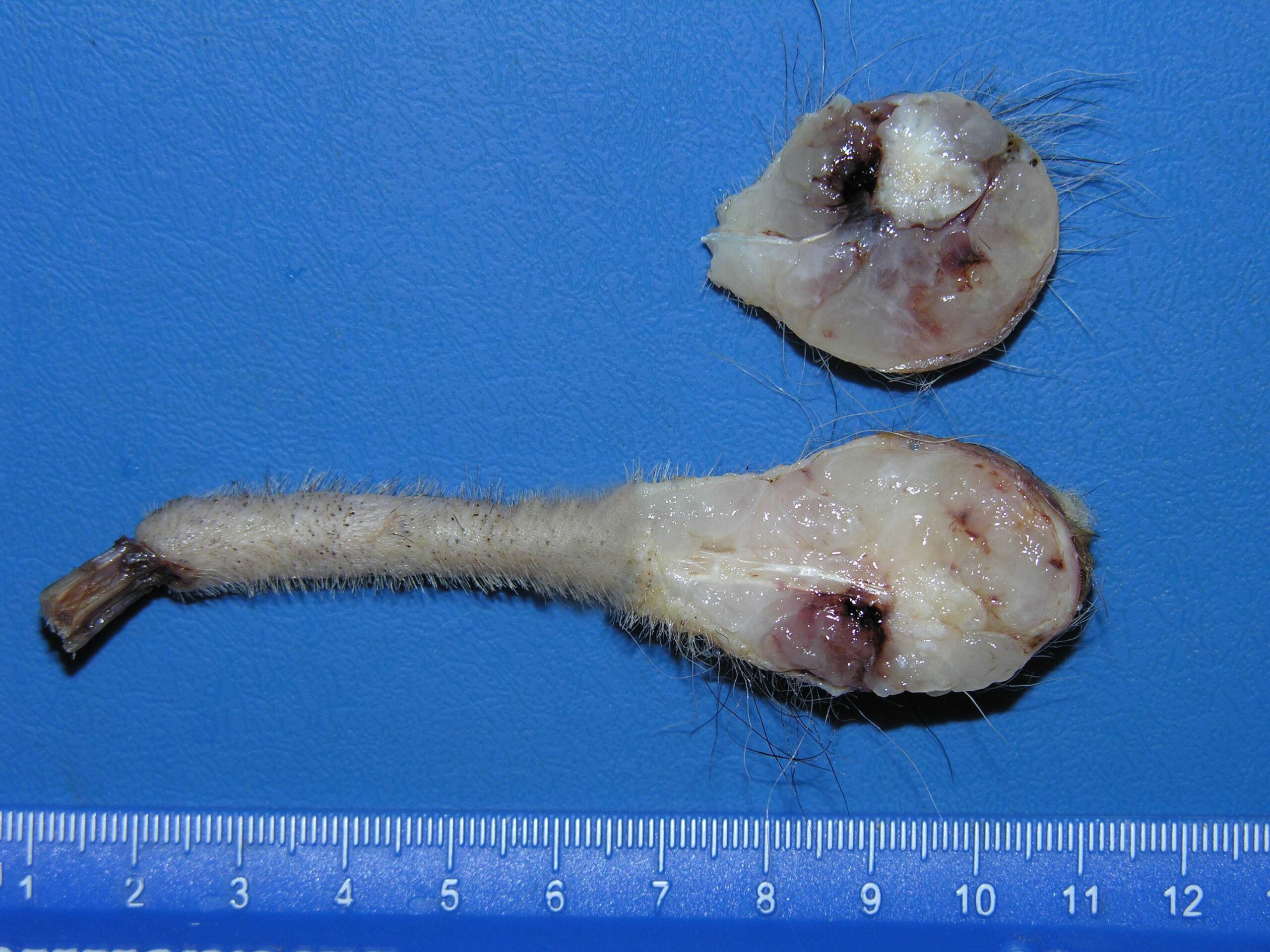
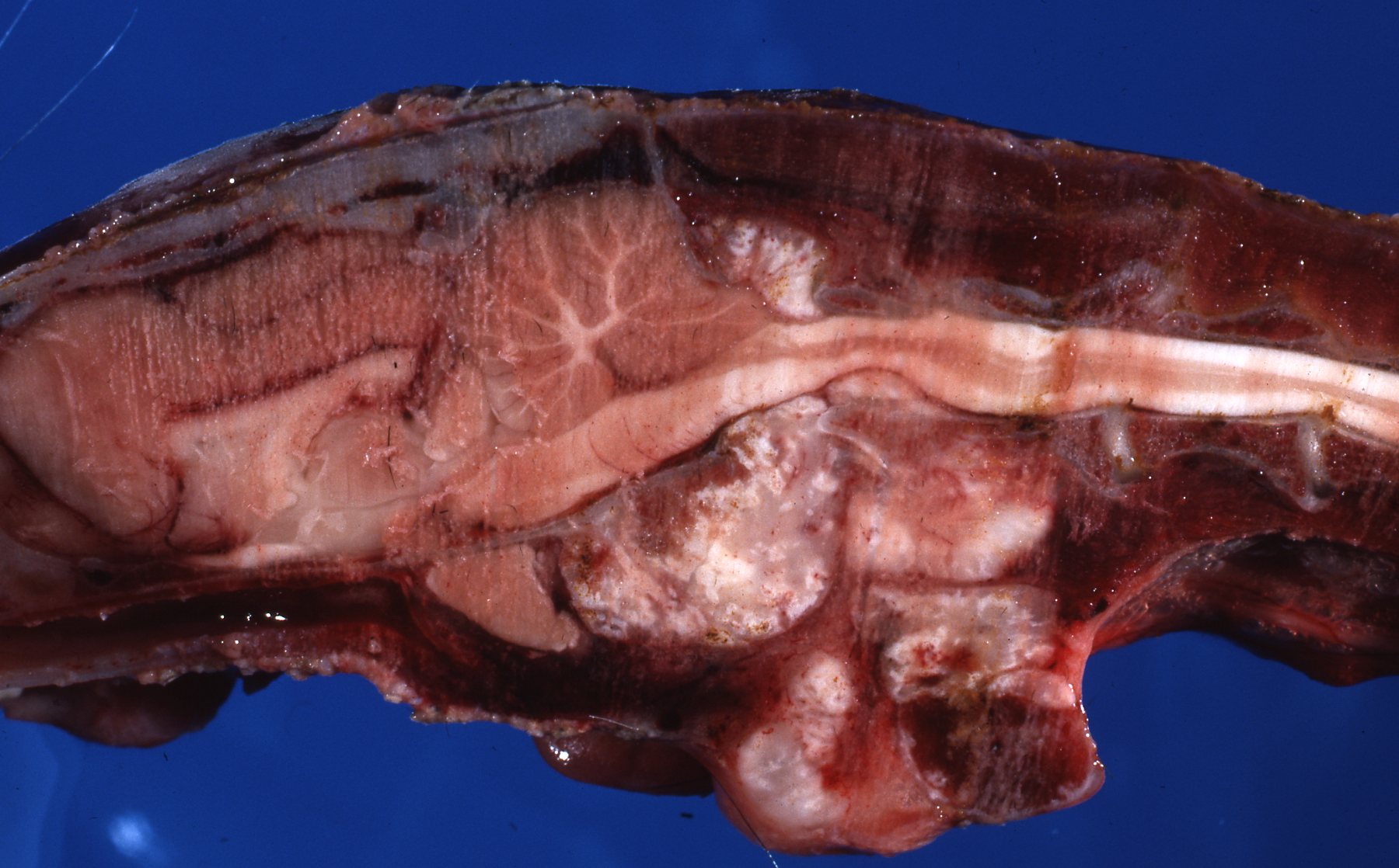
𝐏𝐡𝐨𝐭𝐨𝐬
1-3) Examples of chordomas at the end of ferret tails.
4) An example of a chordoma occurring near the brain, causing compression of the brainstem. Poor ferret!
5) Examples of what a pathologist might see under a microscope! See the lovely bubbles?
6-7) Examples of the same tumour stained for vimentin and cytokeratin to help confirm the diagnosis. I only included these because they look pretty ![]()
𝐒𝐨𝐮𝐫𝐜𝐞𝐬
Maxie, G. Jubb, Kennedy and Palmer’s Pathology of Domestic Animals, Volume 1. Sixth Edition.
Photos 1-7 © Noah’s Arkive contributors Rech, Jakowski, Wilson, Taylor licensed under CC BY-SA 4.0.


