Happy Saturday! This post will be a bit different from our usual content, but I hope you guys find it interesting! Today we are going to discuss a clinical case I had back in 2019 during vet school.
This case is a 16 year old gelding who presented for lameness in both front feet. On physical exam, I noted that his front feet flared out at the ground surface, and the hooves had an extensive ridge pattern on the hoof wall. These changes in the hoof wall are often called laminitic rings, and gives you a clue about what’s going on in this case!
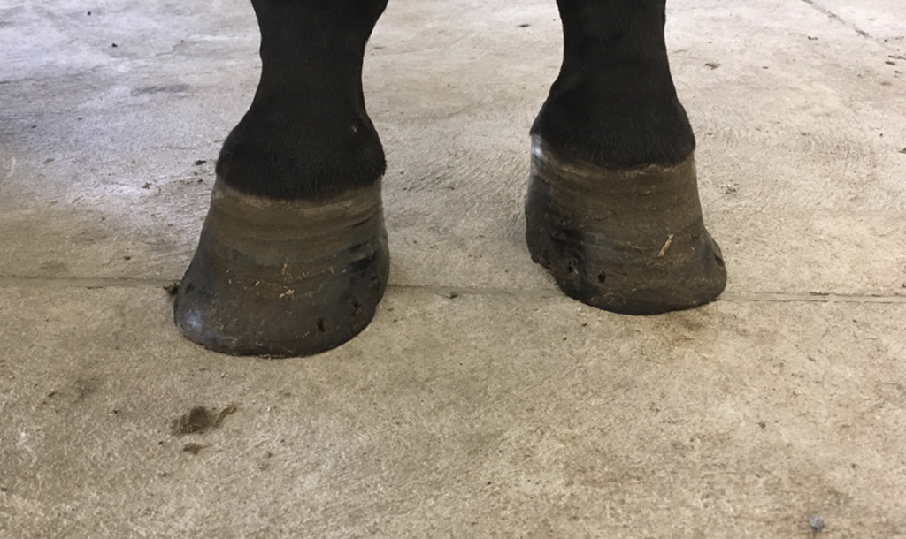
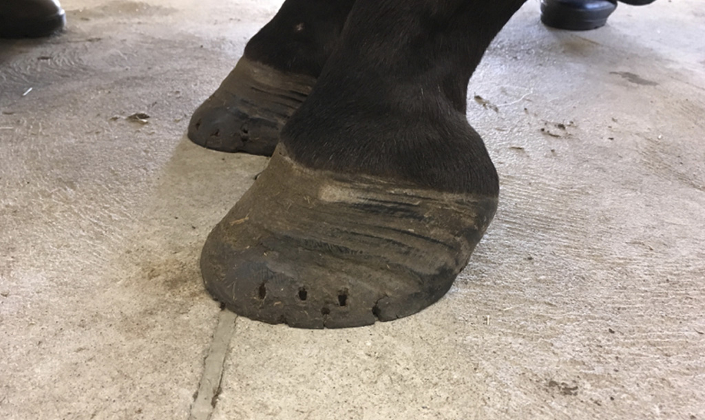
We continued with a thorough lameness exam, and noted that he was most lame on the left front limb. We performed a palmar distal block, which numbs the nerves going to the majority of the foot. With his left front foot numb, it revealed he was also lame on his right front!
So, we moved onto taking X-rays. One of the most striking findings was blunting of the distal phalanx, the bone found within the hoof. Normally, this bone has very straight surfaces with a pointed tip. In this case, the distal phalanx had a significant domed shape on the upper surface, and the tip of the bone was curved upwards like a ski slope. Again, these are characteristic changes associated with chronic laminitis, or laminitis that has been occurring for a long time.

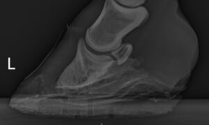
We analyzed the X-rays we took to assess the severity of the laminitis. Arguably the most important measurement is the rotation angle, where the angle of the distal phalanx is compared to the hoof wall. This angle should be 0º in normal horses. In this case, the angle was 4º. Rotation of the phalanx within the hoof is one of the major findings of laminitis, and occurs due to separation of the lamellae. More on this later! We also measured the distal hoof wall distance, or the length between the hoof wall and the phalanx. In normal horses, this should be 15-16mm. In this case, it was 19mm, indicating significant flaring of the hoof wall and rotation of the phalanx. Finally, we measured the founder distance, which is the distance between the top of the hoof and the phalanx. Normally this measurement should be under 4mm, however this case it was 8.5mm! This indicates that not only has the phalanx rotated within the hoof capsule, but it has also sunk downward due to the weight of the horse. Ouch!

So what the heck is laminitis anyway?
As mentioned, laminitis is separation of the lamellae, which are long finger-like projections between the hoof wall and the phalanx. Under normal circumstance, these lamellae keep the hoof wall tightly adhered to the underlying bone. However, certain conditions can cause these fingers to separate from each other, weakening the attachment between the bone and the hoof wall. Because horses are very heavy, this ultimately results in rotation of the bone away from the hoof wall, putting tension on the lamellae. This is incredibly painful for the horse, as these tissues are some of the most sensitive parts of the body. If you’ve ever ripped part of your fingernail off, you’ll know that pain!

So why does laminitis occur?
There are actually several potential causes of laminitis, all resulting from inflammation of the very sensitive lamellae. Chronic laminitis, however, is usually caused by metabolic conditions. In the case of this horse, it was suspected that he had equine metabolic syndrome (EMS), which is sort of like horse diabetes. Horses with EMS have dysfunctional insulin, the main hormone used to move sugar from the blood into the cells. These horses constantly have high levels of insulin, which is thought to affect the function of cells in the lamellae, leading to their separation. Thus, these horses are predisposed to laminitis.
How is it treated?
Equine metabolic syndrome is typically treated by nutritional management and weight loss, since these horses are often quite fat! These horses should be moved off of high-sugar grass and hay, and be provided hay in nets so they cannot take in large mouthfuls at a time. They should also be exercised as much as possible, starting slowly and increasing their workload. However, it is important to remember that laminitis is very painful! If the horse is actively going through a laminitic event, they should be rested and given anti-inflammatory drugs for pain control.
What was the outcome for this horse?
Unfortunately for this guy, he was euthanized due to the severity of laminitis and the amount of care needed to keep him comfortable. He was donated for teaching, and I was able to keep his lower limb bones! I finally mounted them today, hence the inspiration for this post. They need a bit of TLC since they have been sitting in a box for 2+ years, but I am so glad they can finally be used as a teaching specimen!

Patterson-Kane JC, Karikoski NP, McGowan CM. Paradigm shifts in understanding equine laminitis. The Veterinary Journal 2018;231:(33-40).
Redden, R.F. Clinical and radiographic examination of the equine foot. 49th annual convention of the American Association of Equine Practitioners 2003.
Baxter, G.M. Laminitis. Adams and Stashak’s Lameness in Horses 6th ed. Wiley Blackwell 2011.

