Autoimmune diseases occur when there is failure of self-tolerance, leading to the immune system reacting to its own cells. The most studied of these is systemic lupus erythematosus, although many other autoimmune diseases exist.
Table of Contents
Systemic Lupus Erythematosus
Systemic lupus erythematosus (SLE) occurs when there are antibodies directed against many cell components, however the major target are nuclear proteins. Because the antibodies are directed against these nuclear proteins, virtually every cell in the body can be targeted for destruction. This destruction results in formation of antigen-antibody complexes, which can lead to secondary inflammation in the vessels, skin, joints and glomeruli when the complexes deposit in tissue. This condition has been described in many of our veterinary species.
The nuclear proteins that can be targeted in SLE include DNA, histones, RNA-bound proteins and nucleolar antigens. The antibodies can also be directed against red blood cells, platelets, lymphocytes and immunoglobulins. Destruction of these cells can cause anemia, thrombocytopenia and a weakened immune system with leukopenia.
The pathogenesis of SLE is not completely understood, and is likely multifactorial. In humans, it has been shown that MHC genes regulate the production of antinuclear antibodies, which may be similar in other species. Environmental factors like UV light or certain drugs damaging the cells may also contribute to antinuclear antibody production when the cellular antigens are released. It is thought that activation of either B or T lymphocytes may contribute to SLE, with the end result of autoantibody production.
Lesions of SLE
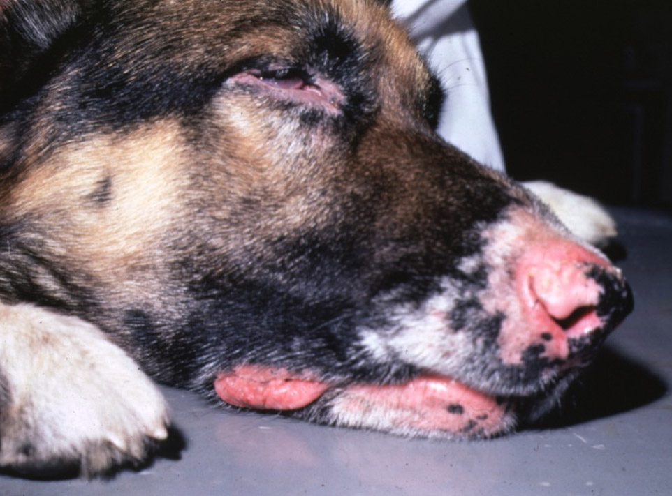
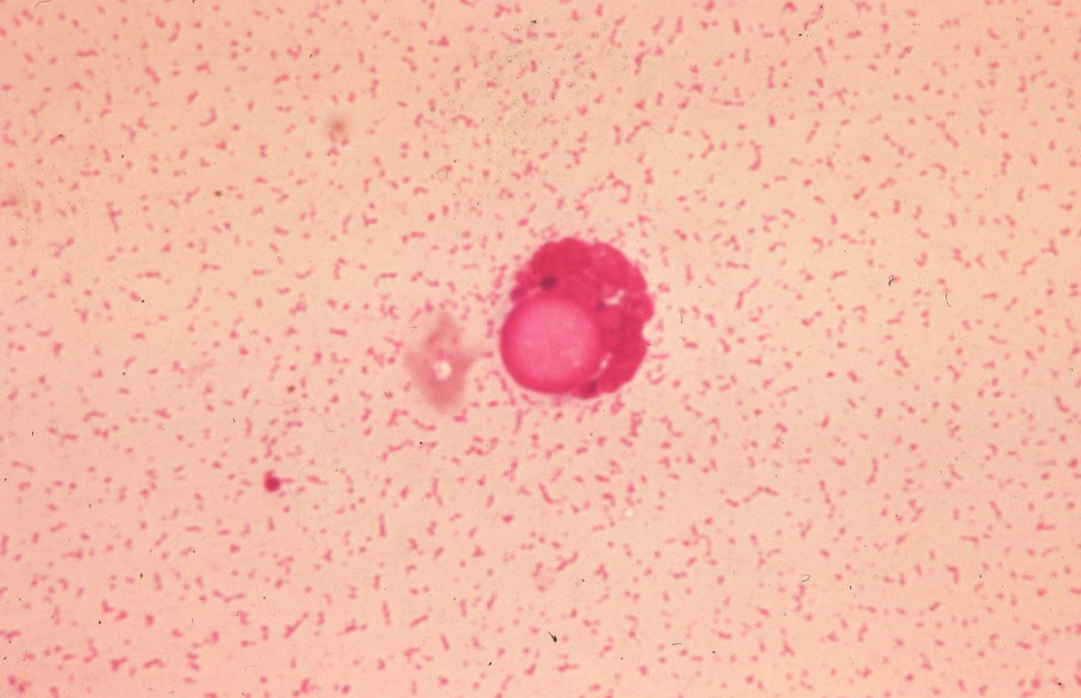
As mentioned, the antigen-antibody complexes depositing in tissue are the main source of SLE lesions. Of these lesions, non-erosive polyarthritis is the most common finding. Histologically, the arthritis inflammation is composed of neutrophils, lymphocytes and plasma cells. Often there is fibrin within the synovium as well. In the kidney, the complexes produce a glomerulonephritis with proliferation. Complexes depositing in the skin produces an ulcerative dermatitis with basal cell vacuolation and necrosis. In any location where the complexes deposit, lupus erythematosus bodies (LE bodies) can be seen, which are eosinophilic cell-free nuclei that have bound to the antinuclear antibodies.

© Vtscharner licensed under CC BY-SA 4.0.

© Latimer licensed under CC BY-SA 4.0.
Rheumatoid Arthritis
Rheumatoid arthritis is characterized by an erosive arthritis, which is caused by antibodies against immunoglobulins. The exact stimulus that causes rheumatoid arthritis is unknown, but may be related to collagen or proteoglycans acting as an antigen. Affected joints often have neutrophilic inflammation and exuberant fibroblast proliferation, called pannus. These fibroblasts may degrade underlying cartilage and prevent synovial fluid flow, resulting in chondrocyte necrosis.
Sjögren-Like Syndrome
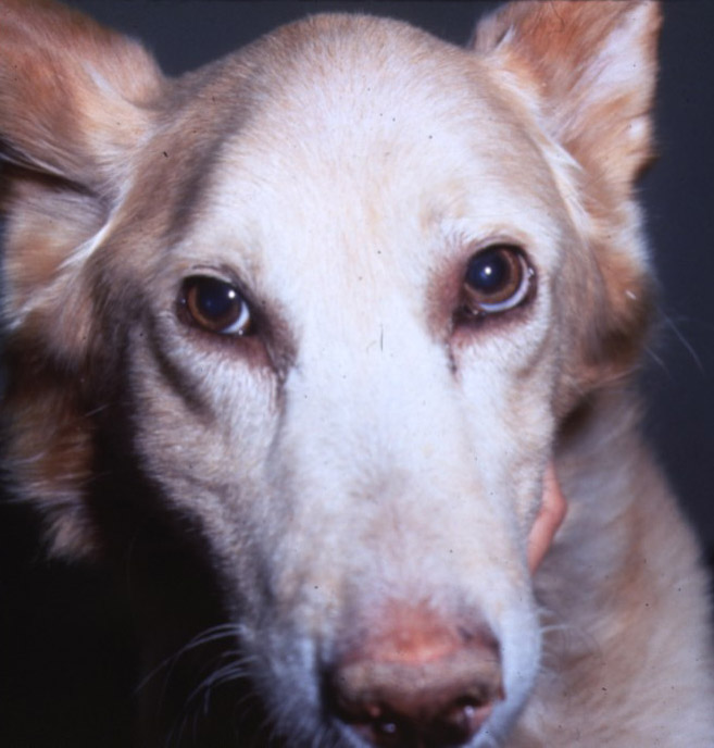
Sjögren-like syndrome is an autoimmune disease that targets secretory glands, particularly the lacrimal and salivary glands. This results in fibrosis of these glands, with lymphoplasmacytic inflammation. Clinically, these animals present with keratoconjunctivitis sicca, xerostomia (dry mouth) and enlarged salivary glands.
Immune-Mediated Myopathies
There are four main myopathies that are thought to be immune-mediated: masticatory myositis, generalized myositis, dermatomyositis and extraocular myositis. All of these present with degeneration of the affected muscle(s), with infiltration of large amounts of neutrophils. In dermatomyositis, the skin is also affected with an erosive dermatitis, similar to SLE.

© Dhein licensed under CC BY-SA 4.0.
Vasculitis
Deposition of antibody-antigen complexes in the vessels produces an inflammatory response, via a type III hypersensitivity mechanism.
One of the major conditions causing immune-mediated vasculitis is beagle pain syndrome, where dogs present with a fever, hunched stance, cervical pain and lameness. The disease is cyclical, with animals seemingly to spontaneously recover, only to have the disease recur. Most dogs will completely recover with no future episodes by the age of 18 months. Histologically, this condition shows necrotizing vasculitis and perivasculitis in multiple tissues. Amyloidosis can also be seen due to AA amyloid accumulation from the inflammatory response.
Zachary JF. Pathologic Basis of Veterinary Disease, Sixth Edition.
Kumar V, Abbas AK, Aster JC. Robbins and Cotran Pathologic Basis of Disease, Tenth Edition.


