Today’s path rounds are on 𝐚𝐦𝐲𝐥𝐨𝐢𝐝𝐨𝐬𝐢𝐬!
𝐖𝐡𝐚𝐭 𝐢𝐬 𝐢𝐭?
𝐀𝐦𝐲𝐥𝐨𝐢𝐝𝐨𝐬𝐢𝐬 is caused by a group of diseases that result in high levels of 𝐚𝐦𝐲𝐥𝐨𝐢𝐝 being deposited in the tissues. Amyloid is a protein material that has a characteristic folding pattern, but has many forms produced in many different situations.
𝐖𝐡𝐨 𝐠𝐞𝐭𝐬 𝐢𝐭?
Any species can get this!
𝐖𝐡𝐚𝐭 𝐜𝐚𝐮𝐬𝐞𝐬 𝐢𝐭?
The most common causes of amyloidosis are inflammation and certain types of tumours. In humans, we also see it associated with Alzheimer’s.
Inflammation causes amyloidosis by producing large amounts of 𝐀𝐀 𝐚𝐦𝐲𝐥𝐨𝐢𝐝, which is derived from 𝐬𝐞𝐫𝐮𝐦 𝐚𝐦𝐲𝐥𝐨𝐢𝐝 𝐀. SAA is an 𝐚𝐜𝐮𝐭𝐞 𝐩𝐡𝐚𝐬𝐞 𝐩𝐫𝐨𝐭𝐞𝐢𝐧, which is a group of proteins produced during inflammation. SAA is broken down in to AA amyloid, so when there is chronic inflammation leading to large amounts of SAA, AA amyloid can accumulate.
𝐀𝐋 𝐚𝐦𝐲𝐥𝐨𝐢𝐝 is produced by tumours of 𝐩𝐥𝐚𝐬𝐦𝐚 𝐜𝐞𝐥𝐥𝐬, which are the cells that produce antibodies. Antibodies are made up of 𝐥𝐢𝐠𝐡𝐭 𝐜𝐡𝐚𝐢𝐧𝐬, so when there is a plasma cell tumour with tons of antibodies produced, these light chains can accumulate in large numbers.
𝐖𝐡𝐲 𝐢𝐬 𝐭𝐡𝐢𝐬 𝐚 𝐩𝐫𝐨𝐛𝐥𝐞𝐦?
Amyloid tends to accumulate in the kidney and liver, causing expansion of these tissues by take up space in the organ. Eventually, this can lead to pressure 𝐧𝐞𝐜𝐫𝐨𝐬𝐢𝐬 (cell death), which often heals by scarring. If enough of the organ is affected, you may see signs of liver failure or kidney failure.
𝐇𝐨𝐰 𝐢𝐬 𝐢𝐭 𝐝𝐢𝐚𝐠𝐧𝐨𝐬𝐞𝐝?
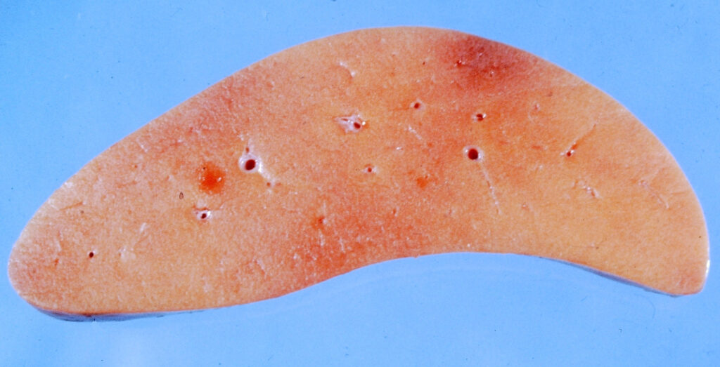
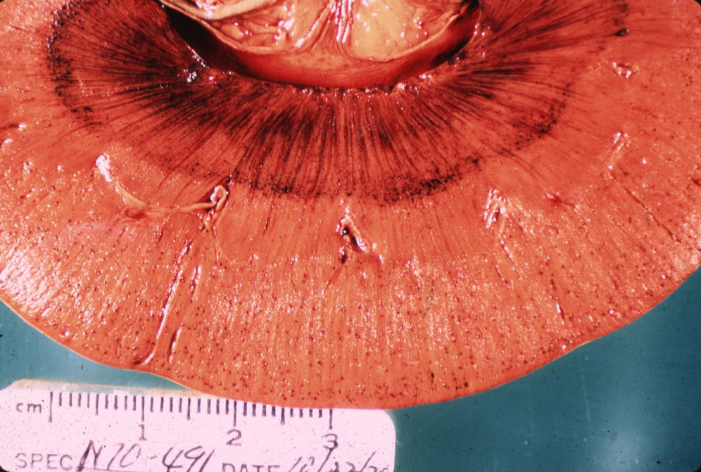
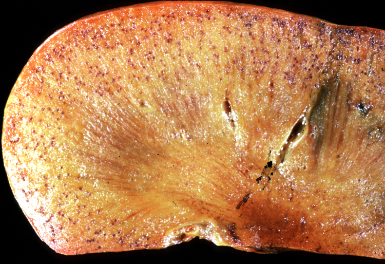
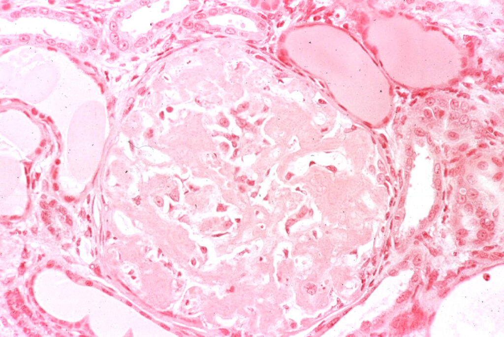
Amyloidosis is typically diagnosed on biopsies or at necropsy. The tissue will generally look quite pale and have a waxy texture, due to the accumulation of the protein. A fun magic trick that pathologists might try is applying 𝐋𝐮𝐠𝐨𝐥’𝐬 𝐬𝐭𝐚𝐢𝐧 to the tissue, which will stain the amyloid dark brown, and make it easy to identify!
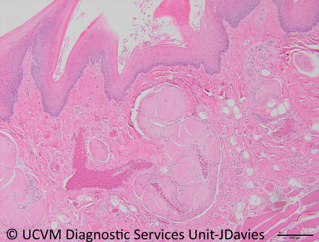
On histology, amyloid has more fun colour changing properties. When a 𝐂𝐨𝐧𝐠𝐨 𝐫𝐞𝐝 stain is applied to the tissue sample, the tissue will glow 𝐚𝐩𝐩𝐥𝐞 𝐠𝐫𝐞𝐞𝐧 under polarized light. Funky!
𝐇𝐨𝐰 𝐢𝐬 𝐢𝐭 𝐭𝐫𝐞𝐚𝐭𝐞𝐝?
Unfortunately there is no specific treatment for amyloidosis, other than identifying and treating the underlying cause.
𝐏𝐡𝐨𝐭𝐨𝐬
1-2) A sample of a kidney and a liver with amyloidosis.
3-4) Kidneys with Lugol’s stain applied!
5-7) What amyloid looks like under our typical stain, H&E, then under Congo Red with normal light, and then under polarized light.
8-9) Another Congo Red and polarized light example!
𝐒𝐨𝐮𝐫𝐜𝐞𝐬
Maxie, G. Jubb, Kennedy and Palmer’s Pathology of Domestic Animals, Volume 2. Sixth Edition.
Photos 1-4, 8-9 © Noah’s Arkive contributors King, McGavin, Read, Williams, Crowell licensed under CC BY-SA 4.0.
Photos 5-7© University of Calgary Diagnostic Services Unit.


