In order for leukocytes to reach an area of inflammation, they must first be able to move out of the bloodstream. This process is called the leukocyte adhesion cascade, and is mediated by the endothelial cells lining the blood vessels. There are five basic steps in this process: margination, rolling, stable adhesion, transmigration and chemotaxis.
Margination
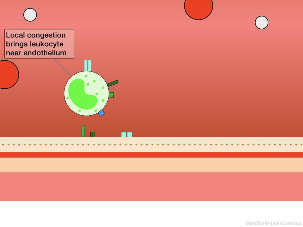
As mentioned in the discussion of endothelia, one of their first responses to inflammatory mediators is to vasodilate and increase the size of the gaps between the cells. Both of these responses cause local congestion in the area of inflammation, with relatively little blood flow. Under normal circumstances, the cellular components of the blood are kept within a central “channel” due to blood velocity, with a thin layer of plasma lining the endothelium and preventing cellular interactions. With congestion, this plasma layer dissipates, and the leukocytes are able to move to the periphery of the vessel lumen. This process is called margination, as the leukocytes are moving to the vessel margins.
Rolling
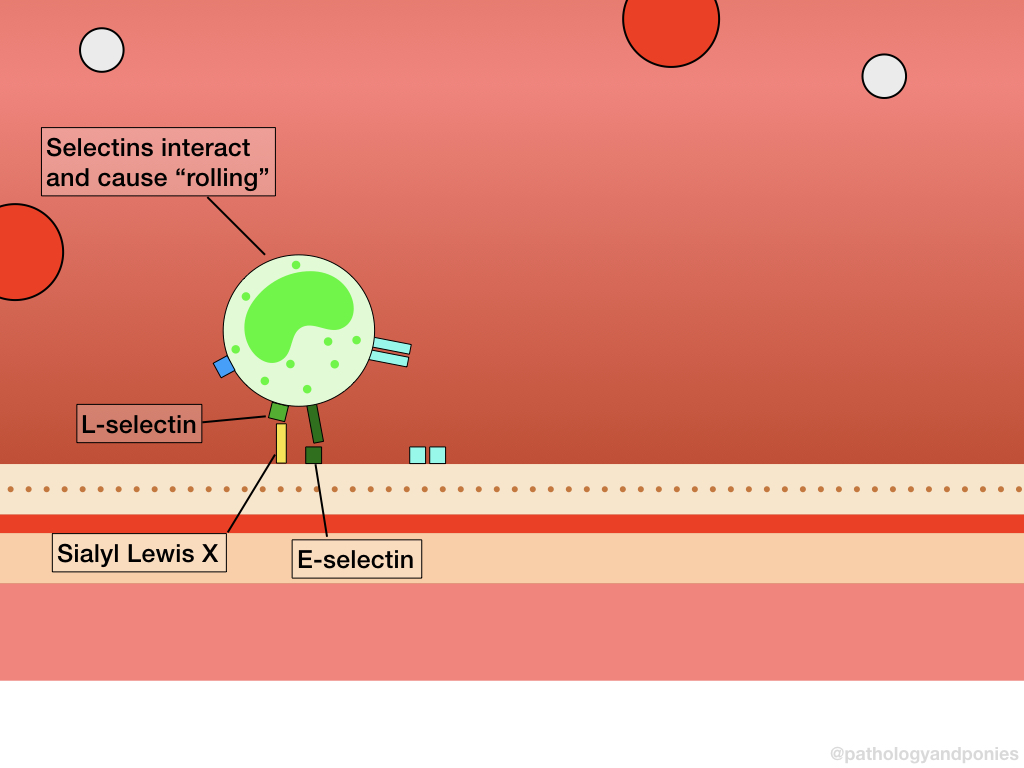
Both leukocytes and endothelial cells activated by inflammatory mediators will express selectins, which mediates the rolling phase of the adhesion cascade. There are three main selectins, which are each expressed by a different cell and bind to different receptors. These are summarized below.
| Selectin | Expressing Cell | Binding Receptor | Cells Expressing Receptor |
|---|---|---|---|
| L-selectin | All Leukocytes | Sialyl Lewis X | Endothelial cells |
| P-selectin | Endothelial cells Platelets | Sialyl Lewis X P-selectin glycoprotein ligand-1 (PSGL-1) | Leukocytes |
| E-selectin | Endothelial cells | Sialyl Lewis X | Leukocytes |
As you can see, the general function of selectins is to attach endothelial cells and leukocytes together. This binding is relatively weak, so these receptors are quickly separated from their ligands by the small amount of blood flow remaining within the vessel. However, this causes the leukocytes to roll, hence the name “rolling” for this step of adhesion. Rolling leukocytes create turbulence in blood flow, reducing local blood velocity even more. This continued process of slowing down the speed of leukocytes eventually allows for stable adhesion, described next.
Stable Adhesion
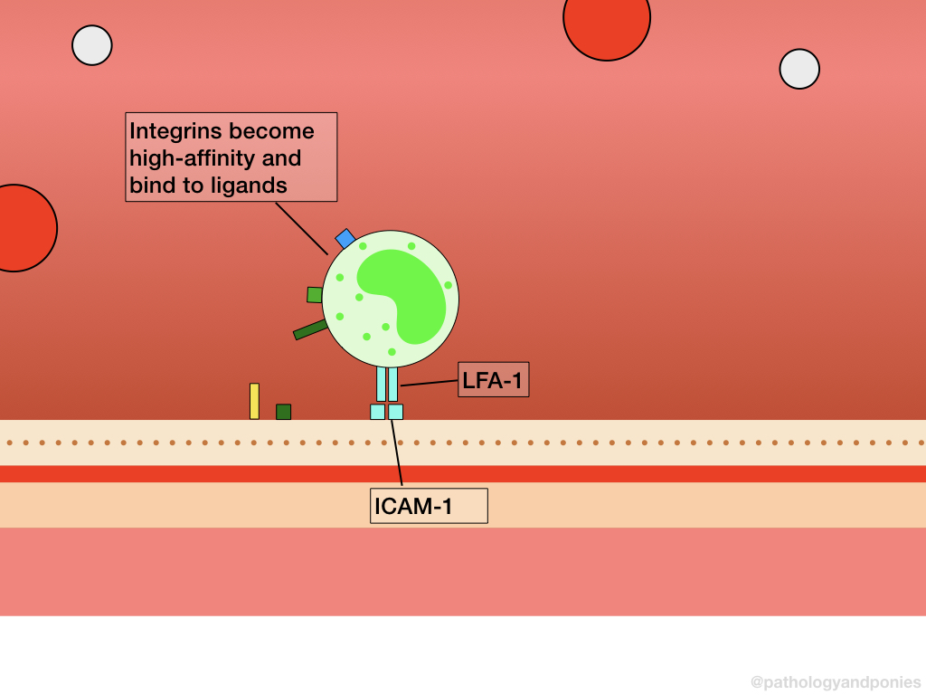
Leukocytes normally express integrins, the major mediators of stable adhesion, however they are usually expressed in a low-affinity state. As inflammation progresses, inflammatory mediators such as IL-1, TNF and C5a will activate leukocytes to cleave their L-selectin molecules from their surface. This cleaving is mediated by ADAM17. After the selectins are removed, the integrins are converted to a high-affinity state, allowing for stable adhesion to proceed. The three major integrins and their ligands are summarized below.
| Integrin | Expressing Cells | Binding Ligand | Cells Expressing Ligand |
|---|---|---|---|
| LFA-1 | Leukocytes | ICAM-1 | Endothelial cells |
| Mac-1 | Leukocytes | ICAM-1 | Endothelial cells |
| VLA-4 | Leukocytes | VCAM-1 | Endothelial cells |
Again, this interaction serves to attach leukocytes to endothelial cells, however this attachment is more secure and not easily washed away. This allows the leukocyte a firm grip to begin transmigration.
Transmigration
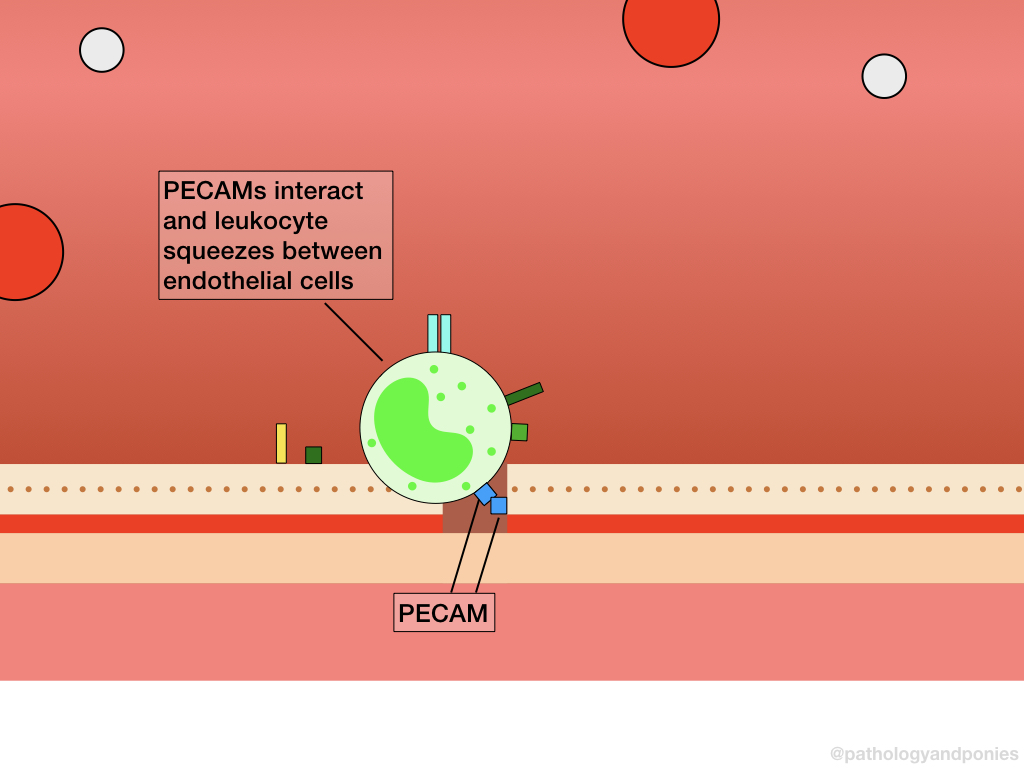
The process of transmigration involves crossing the endothelial wall, and is primarily mediated by PECAM (CD31) and JAMs.
PECAM, otherwise known as CD31, is expressed on both endothelial cells and leukocytes, and binds to itself. Once two PECAMs interact, the leukocyte is able to squeeze between endothelial cells and release enzymes that break down the basement membrane. With the basement membrane out of the way, the leukocyte can continue into the surrounding tissue. JAMs, or junctional adhesion molecules, also facilitate this attachment by binding to integrins on the leukocyte. JAM A can bind to LFA-1, while JAM C binds to Mac-1.
Chemotaxis
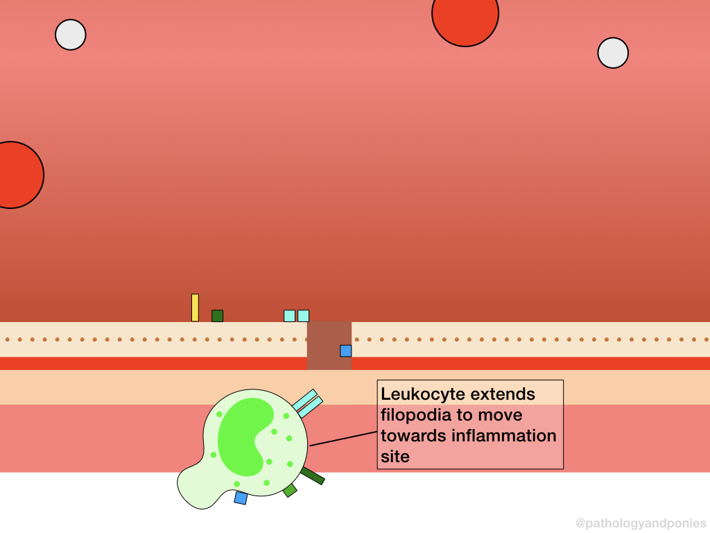
Finally, the leukocyte needs to know where to go! Leukocytes follow a chemotactic gradient, or a gradient of chemoattractants that eventually lead to the source of inflammation. These chemoattractants cacn include bacterial products, inflammatory mediators, cytokines and other metabolites. Because the site of inflammation will have the largest amount of chemoattractants locally, the leukocyte merely needs to move towards an increasing chemokine concentration. The chemoattractant will bind to G-protein coupled receptors on the cell surface, which results in polymerization of actin at the leading edge of the cell and myosin accumulation at the back of the cell. This reorganization allows the leukocyte to extend a filopodia (a foot) towards the higher chemoattractant concentration, to pull the cell along. Eventually, the cell will end up where it is needed. Neat!
Zachary JF. Pathologic Basis of Veterinary Disease, Sixth Edition.
Kumar V, Abbas AK, Aster JC. Robbins and Cotran Pathologic Basis of Disease, Tenth Edition.
Murphy KP, Janeway CA, Travers P et al. Janeway’s Immunobiology, Eighth Edition.

